Exploring the Effectiveness of Distraction Arthroplasty in Joint Rehabilitation

Joint pain, often caused by conditions like arthritis, can significantly impact your mobility and quality of life. While traditional treatments exist, including medication and surgery like joint replacement, a newer technique called distraction arthroplasty is gaining traction. Time to look at what the procedure entails and explore its potential in joint rehabilitation.

What is Distraction Arthroplasty?

Unlike traditional joint replacement, which involves implanting artificial components, distraction arthroplasty focuses on restoring the natural joint. This minimally invasive surgery involves placing an external fixator, a metal frame, around the affected joint. Gradually, over several weeks, the frame separates the joint surfaces, creating a gap. This "distraction" aims to:

  • Unload the joint: By taking pressure off the joint surfaces, this technique promotes the growth of new cartilage and bone tissue.

  • Stimulate healing: The distraction process is believed to trigger the body's natural healing mechanisms, promoting tissue regeneration.

Benefits and Potential of Distraction Arthroplasty

The potential benefits of distraction arthroplasty compared to traditional joint replacement include:

  • Preservation of natural joint: Unlike joint replacement, this surgery aims to heal and restore the natural joint, potentially offering greater long-term function and flexibility.

  • Reduced pain: Studies suggest that distraction arthroplasty can effectively alleviate pain associated with joint conditions like arthritis.

  • Improved mobility: By restoring the joint and reducing pain, this technique can potentially improve range of motion and overall joint function.

Current Research and Considerations

While promising, distraction arthroplasty is still considered a relatively new technique. More research is needed to fully understand its long-term effectiveness and compare it to other treatment options. But there are many studies which use distraction arthroplasty which can show the advantages of this treatment.

Conclusion

Distraction arthroplasty offers a potentially revolutionary approach to joint rehabilitation. While further research is necessary, the potential to restore the natural joint and alleviate pain makes it a promising option for individuals struggling with joint conditions. If you're exploring treatment options for joint pain, it's crucial to consult with a qualified healthcare professional to discuss your specific situation and determine the most suitable approach for your individual needs.

Limb Lengthening and Reconstruction Surgery: What You Need to Know

Limb lengthening and reconstruction surgery is a specialized procedure that can help improve the function and appearance of limbs that have been affected by a variety of conditions. These conditions may include:

Leg Length Discrepancy (LLD)

Treatment goals are to correct the LLD while preserving muscle and join functions and restoring normal limb alignment.

Post-traumatic Bone deformity

Traumatic fractures that heal in displaced, angulated, rotated, or shortened positions.

Non/malunions

Nonunion are unhealing fractures, malunion are incorrectly healed fractures.

Bone loss from tumour, trauma, or infection

Bone loss may be a bone defect or as limb length discrepancy or both.

Joint contractures

Limited movement of tight muscles or capsules or scarring of the joint.

Osteomyelitis

Results from fractures or blood borne infection. An effective solution is to resect the abnormal bone and use Ilizarov technique to treat bone defect.

The surgery can be performed on people of all ages, and it can be a life-changing procedure for those who have been struggling with the limitations caused by their condition.

The Process of Limb Lengthening and Reconstruction Surgery

The process of limb lengthening and reconstruction surgery typically involves several steps:

  • Preoperative consultation: During this consultation, the surgeon will discuss your medical history, your condition, and your goals for surgery. They will also perform a physical examination and may order imaging tests, such as X-rays or CT scans.

  • The surgical procedure: The specific details of the surgical procedure will vary depending on the individual patient's needs. However, in general, the surgery will involve making an incision in the leg, and then inserting a device that will gradually lengthen the bone. The device will be adjusted over time, and the bone will slowly grow new bone tissue to fill in the gap.

  • Post-operative care: In a standard patient, they must remain in hospital for 3-7 days before discharge. Following there is the adjustment and consolidation phase. The adjustment phase is split to 2 further phases:

    • Latent Phase:

For restoration of endosteal and periosteal blood vessels

    • Distraction Phase:

1mm/day until desired length is achieved.

After the desired length is achieved and the limb is straightened, the apparatus is left for the consolidation phase which allows the bone to harden and mature. Once the bone is sufficiently strong, the frame can be removed. The patient is seen every month within these phases once the frame is off with no complications the patient can then have follow-up appointments to the discretion of the Doctor.

Potential Complications of Limb Lengthening and Reconstruction Surgery

As with any surgery, there are potential complications associated with limb lengthening and reconstruction surgery. These complications may include:

Anaesthetic complications

Infection

Deep infections are rare; however, pin tract infections are common but easily treated with pin tract cleaning and oral antibiotics.

DVT/Pulmonary Embolus

DVT is a clot in the leg that can break off to the lung to where it is called pulmonary embolus. Number of precautions are taken, including using a blood thinner called Clexane and the patient is to be blood tested regularly until clear.

Nerve/Blood Vessel injury

Despite being extremely rare, still must account for the risk. Injury of the nerve can cause momentary/permanent partial or total loss of function. Blood vessel injuries may cause significant blood loss, leading to ischemia of a limb.

Non/Malunion

Poor bone formation can lead to nonunion, where the bone fails to heal. But when the bone heals in an unsatisfactory position, that is malunion.

Fracture/Late deformity

May happen due to removal of the frame being too early

Over/Underlengthening

                       Possible, but very rare due to the Doctor being in control of this

CRPS

The nerves act hyper actively and sensitively. Characterised by severe pain, swelling, and stiffness. Unpredictable in nature unless there is prior history.

Loss of function

Reference: O’Carrigan, T. (2016, August 1). Limb Lengthening and Reconstruction. Limb Reconstruction Service NSW. https://www.timocarrigan.com.au/limb-lengthening-and-reconstruction-specialist-orthopaedic-surgeon-campbelltown-nsw.html

Disclaimer: This blog is for informational purposes only and should not be considered medical advice. Please consult with your healthcare provider for any questions or concerns regarding your individual health.

Stem Cells in Arthritis

Author: Dr. Gordon Slater

Arthritis is a widespread joint disorder that can lead to debilitating pain, inflammation, and decreased mobility. While various treatments are available to manage its symptoms, regenerative medicine techniques such as stem cell therapy have gained attention as a potential game-changer in musculoskeletal medicine.

In this article, we delve into stem cells in arthritis, exploring what therapy entails, how it is performed, and who is the ideal candidate.


What are Stem Cells?

Stem cell therapy, a regenerative medicine approach, uses stem cells to promote tissue repair in the body. Stem cells are essential building block cells with the unique ability to develop into various other cell types. This makes them invaluable in restoring damaged or degenerated tissues, including joint cartilage, in cases of arthritis.

Stem Cells in Arthritis

In the context of arthritis, stem cell therapy typically involves using mesenchymal stem cells (MSCs). These MSCs can be harvested from different sources, such as the patient's bone marrow and adipose tissue. Once collected, these stem cells are processed and prepared for injection into the affected joint.


How is Stem Cell Therapy Performed?

The process of stem cell therapy in arthritis can be summarized in several key steps:

1. Patient Evaluation: An experienced healthcare provider, such as an orthopedic surgeon, assesses the patient's medical history, conducts a physical examination, and may use imaging studies to determine the severity of joint damage.


2. Stem Cell Harvesting: The selected source for stem cells, whether bone marrow or adipose tissue is harvested using minimally invasive techniques.


3. Processing and Concentration: The collected stem cells are processed in a laboratory to isolate and concentrate the desired cell population. This concentration step enhances the therapeutic potential of the stem cells.


4. Injection into the Affected Joint: The concentrated stem cell solution is injected directly into the affected joint under sterile conditions, often guided by imaging techniques like ultrasound or fluoroscopy.


5. Recovery and Rehabilitation: Following the procedure, patients typically need to undergo a period of rehabilitative physical therapy to optimize the healing process and improve joint function.


Who Is Recommended for Stem Cell Therapy in Arthritis?

Stem cell therapy is recommended for individuals diagnosed with arthritis, particularly osteoarthritis, where cartilage damage is a primary concern.

Moreover, stem cell therapy may be a suitable choice for:

· Patients with an active lifestyle looking to avoid long recovery periods

· Those who prefer a non-surgical or minimally invasive option

· Individuals who experience persistent joint pain or stiffness

· Patients who have not responded well to conservative, non-invasive therapies such as pain medications, physical therapy, or corticosteroid injections


Side Effects and Drawbacks of Stem Cells in Arthritis

While stem cell therapy holds significant promise, it's essential to acknowledge potential side effects and limitations. As with any injection-based procedure, there are risks of infection at the injection sites. Other drawbacks include:

· Variable effectiveness as results depends on factors such as the patient's age, overall health, and the severity of joint damage.

· Stem cell therapy can be expensive, and the costs may not be covered by medical insurance in some cases.

· Stem cell treatment is not uniformly regulated, leading to variations in treatment quality and outcomes.

· The long-term benefits of stem cells in arthritis are still under investigation, and more research is needed to establish its lasting effects.


Who Is the Ideal Candidate for Stem Cells in Arthritis?

The ideal candidate for stem cell therapy in arthritis typically has early to moderate-stage arthritis with significant joint pain and functional impairment. Additionally, suitable patients are in good overall health, without severe medical conditions that might affect the treatment's safety or efficacy.

Furthermore, they understand the potential benefits and limitations of the therapy, are willing to commit to post-treatment rehabilitation and follow-up care, and have realistic expectations about the outcomes.

Conclusion

Stem cells in arthritis represent a promising avenue for managing pain and overcoming the restrictions associated with this joint condition. As research continues, this innovative approach evolves, providing better outcomes for people with arthritis.

However, while this regenerative medicine technique offers potential benefits, it has side effects and limitations. Moreover, outcomes may vary from patient to patient as it depends on numerous factors, including the patient's overall health, the severity of arthritis, and the skill of the healthcare professional.

If you think stem cell therapy is suitable for your arthritic condition, consult an experienced orthopedic surgeon who can conduct an accurate assessment and offer a personalized treatment plan to use this innovative technique safely and effectively.

Custom Orthotics And Your Orthopaedic Care

3D Print.jpg

Image Credit: 3DPrint.com

Article Authors: Gordon Slater| Tandose Sambo

“Keeping your body healthy is an expression of gratitude to the whole cosmos – the trees, the clouds, everything.” - Thich Nhat Hanh

Orthopaedic care often involves the utilization of special foot supports, in the process of either alleviating or healing your current conditions. Orthotics are special shoe or heel inserts that are prescribed to support your feet during your treatment. As custom made support mechanisms, the orthotic is one of those things that must be carefully designed to fit your body and its needs. 

Orthotics are prescribed for the treatment of a variety of conditions that will be outlined below. Predominantly, they are incorporated into treatments for foot, leg or back problems. Often incorporated into treatment plans as a means to alleviate pain and discomfort, your orthopaedic surgeon will often prescribe an orthotic for conditions such as [1]: 

  • correcting foot deformities

  • helping the foot or ankle function better

  • providing support to the ankle

  • reducing the risks for further injuries

While orthotics often include more than just foot inserts, most people are familiar with that perspective of the orthotic realm. For patients with more serious foot conditions, customized shoe or heel inserts have to be made specifically for the patient’s feet. Custom orthotics are often prescribed once the off the shelf orthotics aren’t effective enough for the treatment desired. 

Orthopaedic Consultations 

Orthotic devices are often prescribed after your podiatrist goes through the process of examining your feet, and investigating the types of symptoms that you are experiencing. A detailed history of your medical history will be collated, in order to ensure that the appropriate treatment plan is created. The medical examination will be one that will assess your feet for any deformities, and pinpoint the sites of pain. 

A series of exercises will also be conducted, in order to identify your feet and ankle positions as you undergo the process of applying loads to them. The pressure distribution of your feet will be determined via imaging technology, and from this data, your orthopaedic surgeon will then be able to determine how to design your orthotic devices. 

For further details your orthopaedic surgeons will also incorporate scans of your feet via technology such as X-rays or an MRI scan. From an external perspective, your feet can only indicate so much, and via the internal checks any soft tissue damage can be determined. From the collective data, the final orthotic device can then be created. 

Orthotic Treatment For Orthopaedic Conditions 

There are a variety of orthopaedic conditions that are treated via orthopaedic custom orthotics. These conditions include: 

  • Arthritis: Osteoarthritis is a degenerative condition that can cause discomfort in the feet. As the gait changes, discomfort in the foot and ankles can result. What orthotic devices will do is provide additional support to the treatment of the condition. With advances in platelet rich plasma technologies, while the joints are healing, the orthotic will alleviate the pain that is associated with the weakened joints. 

  • Bunions. Bunions are painful foot protrusions that do have the ability to cause discomfort during the process of wearing shoes, and also in additional motion. Pre-surgery, orthotics combined with a wide toe box are often incorporated into the shoes, in order to reduce pressure on the big toe. 

This list is not an exhaustive one, just an example of the scope of orthopaedic treatment. Athletes who want to protect their bodies during sporting activities will also incorporate custom orthotics into their preventative strategy.  

Treatment Plan with Orthotics

As a part of a preventative care regime, orthotics are often prescribed along with additional treatments such as physical therapy and anti-inflammatory drugs. As corrective treatments, orthotics really act as braces that the body will then adapt to. Alignment and arch support conditions are almost always treatable with orthotics before surgical treatment will be required. Surgery, as an invasive method, is always the treatment of last resort. 

Types of Orthotic Treatment 

Orthotic devices are very versatile devices. With treatments that can treat spinal, arm and foot and ankle conditions, the prescription for the desired needs is one that will be taken care of. With advancing technology, it is now possible to 3-D print custom orthotics. With the dimensions of the patient known, it will be possible for the orthotic to be printed to fit their requirements. 

The material rigidity is designed based on the intention of healing. In some instances rigid materials such as carbon fiber or plastic will be utilized to make the orthotic. Rigid orthotics are created to provide support.  In some instances, the orthotic may be made from a softer silicone infused orthotic that provides cushioning support. 

Orthotic Design: 

While foot inserts are classified as foot orthotics, they are the simplest form of orthotic that exists. They are usually utilized to treat pain in the feet, and provide cushioning support.  They are available in a multitude of styles including bunion pads and metatarsal pads. 

For escalating pain induced by persistent conditions such as arthritis and flat feet, doctors usually prescribe foot inlays, arch supports and insole. These more advanced orthotics alleviate pain, and optimize patient joint function and gait. 

Once the orthotic is designed, the patient will experience an adjustment period, much like when new shoes are acquired. It takes time for the body to adjust to the new changes. Orthotics are continuously adjusted, in order to ensure that they are fitting the patient.  

Custom orthotics are one of the best ways to keep your feet in great condition if you are experiencing any of the ailments that they are suitable for treating. Talk to your orthopaedic surgeon today, to determine if you should incorporate them into your treatment plan. 

References: 

  1. Healthline : https://www.healthline.com/health/bone-health/orthotics#types

  2. FootCareMD: https://www.footcaremd.org/conditions-treatments/injections-and-other-treatments/orthotics

Gordon+Slater+Logo.jpg

Platelet Rich Plasma And Orthopaedic Care

Alice Pien MD.jpg

Image Credit: Alice Pien, MD 

Article Authors: Gordon Slater| Tandose Sambo 

“Physical fitness is not only one of the most important keys to a healthy body, it is the basis of dynamic and creative intellectual activity.”- John F. Kennedy

In our current medical activities, doctors are now increasingly aware of the mechanisms that enable the human body to heal itself. Regenerative medicine is a field that is facilitating the maximization of the body’s physical ability, and improving the quality of life of patients. With life spans increasing with time, keeping the body in good conditions will be critical to the enjoyment of life. One critical enhancer to human restoration is platelet-rich plasma. This plasma is generated from the body’s own blood supply, and enables the natural growth factors embedded in the plasma to heal relevant tissues in the body. This tissue can include cartilage, a critical component of human joint systems. 

How Is The Plasma Obtained?

The human circulatory system consists of an ever flowing blood stream. The plasma is the liquid portion of the blood. Other constituents that are suspended in the blood include red blood cells, white blood cells, and platelets. The platelet, as a component of the blood, is a type of blood cell that is able to facilitate healing of the body. When there is a cut, the platelets are what accumulate in order to generate the clot, that finally heals into the renewed skin. The significance of the platelets then, cannot be underestimated. The platelet activation process is key to the body’s healing mechanisms. The protein growth factors are also embedded in the platelet structure. 

Via centrifugation extractive processes, the plasma (known as protein rich plasma or PRP) can be obtained. With a higher concentration of platelets than the blood, the healing abilities of the PRP can be optimized from 5 to 10 times the normal healing mechanisms. The healing process is therefore catalyzed. 

Mechanism of Activation

With the PRP extracted from the patient’s blood, the extracted components are then injected into the sites of healing, in order to accelerate the healing of those sites. For patients who are experiencing injuries in their tendons, ligaments, muscles and joints, PRP injections are one of the primary methods that the therapy can be applied to heal the body. Many athletes are actually the recipients of PRP injections, and they have proven to be very effective in the process of restoring musculoskeletal problems. 

According to the severity of your condition, PRP injection treatments may take up to one or several tubes of your platelets. When applied to the body’s injured tissues, the growth factors then start the process of catalyzing healing, and increasing the production of the body’s healing mechanisms. 

During the process of administering the injection, ultrasound imaging is often utilized in order to gain a picture of what is happening in the internal system. With the ultrasound image, your orthopaedic surgeon will be able to see directly into the tendon, and facilitate the guidance of the plasma to the appropriate site in the joint. A variety of ailments have been relieved via the PRP mechanism, particularly for persons with conditions such as tendonitis. 

With the ability of the body to heal itself naturally, the utilization of medication such as anti-inflammatory agents or opioids can be eradicated. As an agent that is produced by your own body, the PRP is one product that your body will readily accept because it is natural to your own system.

Factors That Influence PRP Administration 

PRP effectiveness is currently being studied for a more quantifiable effect on the patients that are treated. The current effectiveness is a variable parameter that is currently being investigated. In terms of studies that are currently being focused on, the parameters of focus include: 

  1. Area of the injured site

  2. Patient Health and Physical Fitness 

  3. Extent of the Injury 

In some instances, PRP has found utility during surgical processes, in order to heal tissues post surgery. During shoulder surgeries, patients with torn rotator cuff tendons are now being injected with PRP. Surgical procedures with the treatment of anterior cruciate ligament are also currently being investigated. 

Talk to your orthopaedic surgeon, if you feel that PRP therapy will be beneficial to your healing. 

Article References: 

  1. HSS: Platelet Rich Plasma: https://bit.ly/3dPGS3f

  2. Orthoinfo: https://orthoinfo.aaos.org/en/treatment/platelet-rich-plasma-prp

Gordon+Slater+Logo.jpg

Stem Cell Structure Co-Cultures And Orthopaedic Care

tissue_engineering.jpg

Image Credits: The Stem Cellar  

Article Authors: Gordon Slater|Tandose Sambo 

“Health is a large word. It embraces not the body only, but the mind and spirit as well; …and not today’s pain or pleasure alone, but the whole being and outlook of a man.”- James H. West

The future of medicine is in stem cells and regenerative therapies. With the utilization of stem cells becoming more prevalent in the medical realms since their discovery and introduction to medical research, the advances in medical research are taking note of the benefits of the therapy. 

One of the areas of medical advancement that has benefited from stem cell therapies is the process of tissue engineering and regeneration. The ability to successfully engineer various tissues is one that is becoming increasingly more beneficial to medical treatment. The therapy is still in its relative infancy, and limitations of stem cell science are being overcome by the utilization of a combination of stem cells with co-culture systems. Stem cells do have regenerative abilities, and the potential to regenerate into differentiated cells according to the environment that they are placed in. They are the starting point for life, and cell differentiation that leads to organism development. 

When stem cells are applied in co-cultures, they promote tissue growth and repair via a multi-fold approach. One of the influences is that of an enhancer in the culture that they are introduced to, via enhancing the existent terminally differentiated cells. In an instance where the cells are few in number, the stem cells will actually facilitate the proliferation of more of those types of cells, and increase cell survival, proliferation, phenotype maintenance and organization. 

Stem cells have a multitude of properties, and have the ability to regulate cell functions within living systems. They assist with the regulation of the co-culture that they are integrated with, via providing a regulatory control loop that enables the system to actually sustain itself, and heal, restore and repair where the system needs to. Via the synergistic operation of the stem cells with their co-culture cells, the stem cells have the ability to act in supporting roles to the system. As their functions are diverse, where they are placed, the stem cells will quickly adapt. 

The design of a co-culture is one that aims to optimize cellular interactions, and improve a biological system. The system could be one that is of a human system, or in some instances in an animal system. The anticipated interactions will be via direct cell–cell contact, cell–ECM adhesion and transfer of signalling molecules[1]. These interactions, while detected by science, are actually still in the process of being deeply understood, in terms of their mechanisms of action. 

The interaction of stem cells and existent terminally differentiated cells in a co-culture is one that is being investigated. What is known is that they do communicate via a mechanism that is still being elucidated. The primary elements of the communication paths include interaction via growth factor dose and dosing regimens. Additional factors within the extracellular matrix (ECM) have also been found to influence the extent of communication between the stem cells and their co-culture counterparts. The ECM influence on the organism is one that is much like larger organisms and their environment. The more nourishing and nurturing the environment, the more likely that the stem cells will maintain and regulate the biological functions of the systems that they are regulating. 

As the tissue engineering and engineering realms understand the relevant factors that lead to improvements in co-culture regulation, the more success there will be in vivo transfers of the system. Tissue engineering is a novel activity that will generate many benefits once it is mastered. The interaction of stem cells and terminally differentiated cells is an engineered environment that doesn’t take place within the body. As an experiment that is geared towards looking at all possibilities, the notion that knowing what happens in an extreme condition, will ready the scientists to understand what will happen in the body is currently being applied. The test conditions will generate the conditions that will ensure that the real conditions will be more readily understood once they are attempted. 

Orthopaedic Soft Tissues

With co-culture conditions generating favorable results, the next steps would involve the process of actually utilizing them in the regeneration of various cells across the body. Within the body, cartilage is one of those systems that takes a very long time to heal itself once it has lost some of its cells due to damage via sporting activity, or via degenerative processes such as osteoarthritis. 

Cartilage as a cellular system is one that is monocellular in nature, and it is generally one that has few vessels and nerves. The nature of cartilage is one that makes it so difficult to regenerate. Systems that are highly vascularized tend to be better able to regenerate themselves because of the access of blood flow and nutrients to the area. The nature of cartilage, therefore makes it one of the more challenging locations to utilize as a cell source for tissue engineering. 

Regenerating the area, therefore becomes one of the biomedical grand challenges. The utilization of stem cells to regenerate cartilage in vivo has proven to be successful. Via stem cell injections into an in vivo site, the ability to increase joint integrity was actually witnessed. After an incubation period, the injected stem cells enabled the body to regenerate itself, and the final results of higher expression of chondrogenic genes, and the improved mechanical integrity of the joints were noticed with time. The results proved the ability of stem cells to produce collagen types I and II, and to optimize the functions of the joint from a mechanical standpoint. Engineered cartilage is gaining traction in medical therapies. Patients who experience conditions such as osteoarthritis, will be the prime candidates for this therapy. 

A Promising Future 

Another future possibility is the fact that co-culture applications are now finding application in multiple spheres. With the ability to utilize more than two cell populations in a single system co-culture, the prospects of increased medical understanding of these systems are becoming better understood. Advantages of this multi-cell co-culture include the ability to heal systems such as cardiac and vascular tissue. In research conditions, conditions such as ischaemia, responded well to the introduction of a cocktail of stem cell types. 

As the ability of co-culture systems to regenerate native tissues expands, they are becoming a gold standard in the tissue engineering of complex tissue. The next phases of the exercise is one that will involve the integration of technologies such as bio-printing, with co-culture technology. As regenerative systems that can build tissues, and ultimately organs, the ability to replace damaged tissues will be a possibility in the future. 

Stem cell co-culture and their applications have shown great promise in their testing conditions, and also in their applications. As they are utilized in tissue engineering applications, the ability to regenerate systems will be enhanced. As a patient, one of the things that you can feel confident about is the possibilities that exist for your healing. Research where you can about whether you feel that stem cell therapies are right for you, and consider them as part of your current or future therapies. 

Reference: 

[1] Advances in tissue engineering through stem cell-based co-culture: https://onlinelibrary.wiley.com/doi/epdf/10.1002/term.1870

Gordon+Slater+Logo.jpg

The Role of Platelet Rich Plasma in Cartilage Repair

Stubbs Dental.jpg

Image Credit: Stubbs Dental

Article Authors: Gordon Slater| Tandose Sambo 

“Our greatest happiness does not depend on the condition of life in which chance has placed us, but is always the result of a good conscience, good health, occupation, and freedom in all just pursuits.” - Thomas Jefferson

Medical science, like all other sciences, is an evolving field. Within the fields of orthopaedics and sports medicine, traditional medicinal practices are being utilized along with more unconventional methods of treatment, that utilize the body’s own mechanisms to optimize the healing of an individual. As the potential of biologics continues to emerge, the future of the utilization of these therapies will continue to dominate. One such therapy is the utilization of platelet rich plasma (PRP). The applications of PRP are starting to emerge within the orthopaedic realm. As an enhancer of cartilage repair and regeneration, its utilization is becoming a standard in orthopaedic care. 

While initial phases of the understanding of the mechanisms of action of PRP were somewhat unknown, there is a current emergence of the various interactions that enable PRP to achieve its objectives of healing and regeneration in a human biological system. As higher quality information is available, the proof of PRP effectiveness is actually being validated. Studies focused on this report are those dedicated to the understanding of the efficacy of PRP in the treatment of articular cartilage injury. This injury is one of those conditions that is identified during the degenerative process that is induced by osteoarthritis (OA). 

During the progression of OA, there are various mechanisms at play that result in the altering of various biological parameters. With these internal changes, the relevant regulation of compounds such as cytokines and various enzymes is affected. PRP as a treatment is one that is able to counteract these conditions, and ultimately result in the restoration of the appropriate balance of these internal factors, and enable the joint to sustain itself and restore. As pre-clinical studies and clinical trials are optimized in terms of technology and focus, the effectiveness of PRP and its direct mechanisms of action will be well known.

OSTEOARTHRITIS

Osteoarthritis as a condition is one that affects the knees mostly, and is attributed to the loss of articular cartilage. Affecting up to 4% of the globe at any one time, there is a need to ensure that the healing of this condition can be optimized. With longevity on the rise, mobility of the human population will be one thing that persons will want to factor into their quality of life. Biologic utilization in the treatment of OA aims to minimize the inflammatory processes that take place during arthritis, that cause the articular cartilage to be affected. To ensure that articular cartilage is restored, it will be important to ensure that critical parameters are sustained. These are the anabolic and catabolic factors. PRP does well in the balancing of these two. 

PRP IMPACT ON OA 

PRP is obtained via centrifugal processes, and involves the extraction of the platelets from the bloodstream. Within these concentrated solutions of PRP, are the various healing components that will facilitate the restoration of the articular cartilage. This includes the increased concentration of growth factors and proteins. When concentrated in a location such as the knee, and injected to a healing site, these components will then enhance the regeneration of the tissues. Articular cartilage is degenerated in the OA progression, while PRP counteracts its mechanism of action via: inhibiting the catabolic cytokines of IL-1β and TNF-α, and by promoting factors associated with cartilage matrix synthesis including fibroblast growth factor, transforming growth factor-β (TGF-β), and others.

As a modulator of the inflammatory processes, PRP has proven its effectiveness in controlling the process. Further details of the mechanisms of action are outlined in the second reference of this article. The process is a very detailed multi-step regulatory process that involves a series of interactions between the PRP and various elements in the cellular matrix. 

PRP is incorporated into a multitude of treatment strategies in the treatment of OA. These include operational utilization via grafting mechanism, or the incorporation of the treatment in a non-operative manner via injections. As the demand for biologic treatment increases, the PRP methodology will be a factor that will be increasingly utilized in the minimization of the impacts of OA. As a regenerative therapy, it will be one of those treatments that has dual purposes. Various pharmaceutical preparations modes of PRP are under study, and the findings and efficacy being documented for future use. These include preparations such as fibrin glue or gel, inactivated leukocyte-poor PRP, and leukocyte-rich PRP. The preparations have been tested in both a pre-clinical and clinical application. 

Clinical Applications of PRP have been a medical focus for the past twenty years and continue to be a benefit to the treatment of OA. As varieties of PRP preparations have become standard and are proving their efficacy, the utilization will only go up. As PRP aims to restore the articular cartilage via mitigating and balancing the inflammatory and catabolic environments in a site, the hope is that a deeper understanding of how this occurs will be finalized during studies. 

The future of PRP utilization is definitely one that will be a focus for many more decades to come. Once the establishment of robust clinical trials and improvement in reporting standards is established, the appropriate escalation to clinical trials will be facilitated. 

Article References: 

  1. The Role of Platelet-Rich Plasma in Cartilage Pathology: An Updated Systematic Review of the Basic Science Evidence: https://bit.ly/3lugXAC

  2. Platelet-Rich Plasma and Cartilage Repair: https://bit.ly/3nDJx4

Gordon+Slater+Logo.jpg

Bone Grafts And Bone Substitutes

person-holding-small-anatomical-model-of-skeleton.jpg

Image Credit: Medical News Today 

"Our health is something we often take for granted. But, there are some things in life that should never be taken for granted. Take care of yourself." Catherine Pulsifer

The bones, and our skeletal structure, are aspects of our bodies that we often don’t take notice of because they are within our internal structure. In some instances, there are a multitude of possibilities that result in the need for bones to be corrected. During these corrective procedures, there are currently a series of bone inclusions and additions that orthopaedic surgeons will utilize in the process of healing the bone structure, and improving the structural integrity of the bones. These options include the utilization of various bone grafts that are either taken from the patient's body, a medically approved cadaver, or even materials that have been fabricated by material scientists. 

With many advances in fields like synthetic bone substitutes, it is quite possible that improvements in human orthopaedic surgery will be achieved. The future of orthopaedic healing, includes the utilization of technologies like composite bone grafts, that contain healing agents embedded in a osteoconductive matrix. Embedded in this matrix includes the combination of osteogenic cells and osteoinductive growth factors. With the right nutritious environment in place, the ability for the bone to regenerate itself will take place. With a variety of options for healing, let us delve into a few critical categories of bone grafts and bone substitutes: 

Autografts

Autografts are bone transfusions, that are generated from the patient's own body, and transferred to the relevant section of the body (such as the spine) in order to be fused with the bone that is currently to be healed. There are usually two or more surgeries that are conducted in the process of treatment. This process usually includes the surgery that is dedicated to remove the bone from the base source. This surgery is known as a harvesting surgery and it is utilized in order to generate the bone graft. Harvested bones are usually taken from locations such as the ribs, the iliac crest of the pelvis, and also sections of the spine. Medical advances are always taking place, and the inherent risks that are associated with harvesting and graft transfer, are currently being optimized by the utilization of material science that generates suitable bone substitutes. 

Factors considered in Autograft Utilization 

Within medical practices, the autograft is classified as the gold standard for certain procedures such as spine fusions. With a spine fusion, the appropriate conditions for a solid bone bridge are achieved. The new bone growth is highly favored. The grafted bone also contains osteophytes and other bone growth enhancers that are able to generate new bone growth. The autografts has found favor in the field because there is [1]:

  • Greater chance of fusion success vs. allograft (cadaver bone) and some types of bone graft substitutes

  • No risk of disease transmission (vs. using cadaver bone).

The main disadvantages of using autograft include the risks and possible complications associated with conducting any surgical procedure, e.g.:

  • Surgical wound problems, such as infection

  • Nerve injury (rare)

  • Bleeding (rare)

There is also a limited supply of this type of bone graft and sometimes it needs to be supplemented with some form of bone graft substitute[1].

Bone Substitutes

Bone substitutes are being increasingly utilized in surgery. The number of bone grafts that are achieved via the utilization of bone substitutes is approaching several million each year. With the limitations that are inherent in the utilization of autografts highlighted above, medical science has identified alternate ways to heal bones that are able to overcome these restraints. 

Bone substitutes include elements such as allografts, xenograft substitutes, and ceramic based synthetic bone structures. Of these three bone substitutes, the ceramic based synthetic bone substitutes are the therapies that are widely used in clinical procedures. These ceramic bone substitutes are proving themselves to be effective. In the minimally invasive realm, there is also the possibility to utilize injectable cements. There is still ongoing research that is dedicated to the advancement of the field. 

Incorporation of a Bone Graft

The incorporation of a bone graft is a process that is defined as a multi-step cascade. Once the transfusion surgery is completed, there will be a healing process that enables the graft to be incorporated into the body. The healing process is one that involves the generation of inflammatory cells, along with the movement of the existing mesenchymal stem cells to the site of the graft. These mesenchymal stem cells will then intelligently differentiate into healing cells known as chrondoblasts and osteoblasts, and facilitate the healing of the bone. There is a regeneration of blood flow to the graft site, known as revascularization. Via a series of mechanisms, eventually the bone is internally remodeled, in order to restore the integrity of the bone, and enable the patient to regain full mobility and weight bearing capacity. 

What Is The Ideal Bone Substitute? 

The ideal bone substitute is a composite that is biocompatible to the healing site. In some instances, there have been adverse effects of the incorporation of a bone substitute. The immune system in general is very sensitive to any foreign bodies that are introduced to it, and there is often an immune response that initially attacks, then embraces the graft. The reaction to grafts is an individual one. Biocompatiblity is a capability of new bone substitutes, that will enable the substitute to be incorporated into the body within the minimal amount of time. The key parameters that bone substitutes should embody, are identified by the medical community. When analysis is conducted, these key parameters are to be facilitated by any material that is to be incorporated into the body. The parameters not only factor in the ability for the bone structures to be incorporated into the body, but also for the material to be affordable. 

Advances in technology, will ensure that future treatments are customizeable to the patient’s body. Until such time, the current available pool is one that is increasingly enabling faster patient recovery, and inherent adaptability for inclusion into the body. 

References: 

  1. Spine Health: https://www.spine-health.com/treatment/spinal-fusion/autograft-patients-own-bone

  2. Bone Substitutes in Orthopaedic Surgery: https://bit.ly/30Kg1Aj

Gordon+Slater+Logo.jpg

Foot Ulcers And Their Impact on Diabetic Charcot Foot

ndiabeticfoot.jpg

Image Credit: ndiabeticfoot.org 

Authors: Gordon Slater| Tandose Sambo 

“The secret of health for both mind and body is not to mourn for the past, not to worry about the future, or not to anticipate troubles, but to live in the present moment wisely and earnestly.”- Buddha

What is a Foot Ulcer? 

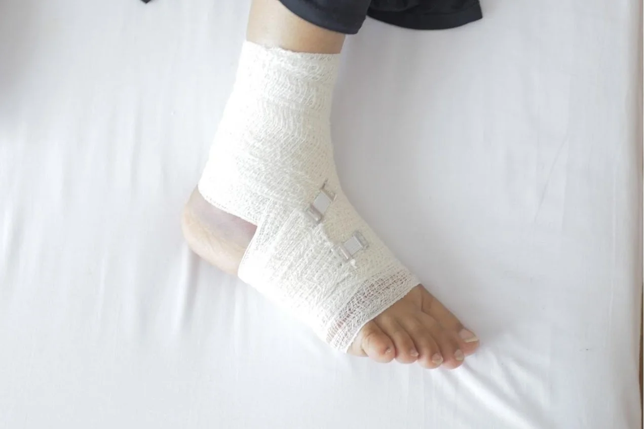
Foot ulcers occur when there’s a deep sore that develops on the foot. With careful care, the condition can be treated and healed, and in the adverse condition it becomes infected. The conditions that induce foot ulcers include cuts from external sources, or even from an abrasive shoe. Foot ulcers are common in diabetics, and if they are not carefully monitored, they can degenerate into adverse conditions that may require either surgery or amputation. 

What is Charcot Foot? 

Charcot Foot is a severe complication of diabetes that ultimately results in the weakening of bones, joints and tissues in the foot and ankle regions. The condition is a downstream effect of the presence of peripheral neuropathy, that causes the patient to lose their ability to feel pain. This condition is classified as an insensate condition. The nervous system is the means via which we are able to sense and feel the environment. Without this capability, patients often experience injuries such as cuts, and are unaware of them unless they take the time to actually inspect their feet on a daily basis. 

As Charcot foot progresses, there is an associated weakening of the bones that causes fractures and joint dislocation. As the joint collapses, there is an eventual deformity of the foot that results from the inability of the joint system to sustain itself. The eventual appearance is a rocker-bottom appearance. Under such conditions, foot ulcers are able to develop. With open sores on the feet, there is also the possibility of bone infections, as bacteria can travel from the skin into the bone structure. 

Charcot foot is one of those conditions that can be mistakenly diagnosed, because it is often the result of some underlying condition such as diabetes. As diabetes is on the rise, the occurrences of Charcot foot are expected to increase in a linear fashion. For this reason, diabetics need to be aware of the fact that they can develop this condition, and take the necessary precautions and care to ensure that their systems are kept under control. Visits to specialists like vascular surgeons and podiatrists will ensure that the adequate foot health is being maintained. 

Causes and Clinical Course

Charcot foot is defined as a neuropathy induced condition which results in the weakening of the ankle joint, and ultimately the flattening of the feet. Neuropathy patients often can’t feel their extremities due to nerve damage, and ultimately end up deforming the foot as the joint collapses. This condition is quite serious, and several treatments have been proposed, that facilitate the restoration of balance in the body. 

Additionally, Charcot foot can develop as a downstream effect of an untreated injury. As a condition that results in the weakening of the ankle joint, if a patient continues to operate with the ankle as it is, the joint will eventually degenerate. As the structure weakens, the joint weakness and subsequent bone loss will result in the collapse of the foot. 

If the foot degeneration is in the middle region of the foot, there will be the formation of the rocker bottom that is associated with Charcot foot. The ankle will eventually become unstable. With Charcot foot, if there are any sharp edges of bone pressure can be placed on the skin from the inside out, and as a result they will ultimately cause the formation of chronic skin sores. If left untreated, these sores can cause foot ulcers, which can be detrimental to the health of the patient. 

Patients should take the time to actually check their feet for indications of any abnormality. This can include changes in the temperature of the feet, redness and swelling, or any occasional soreness that appears to be chronic. 

Diagnosis

The best way to ensure that Charcot foot is diagnosed, is to ensure that the appropriate tests are taken by an Orthopaedic surgeon. The key to ensuring that the foot integrity is maintained, is to ensure that early detection is incorporated into the treatment process. Via medical tests and the utilization of imaging technologies such as X-rays, the appropriate analyses will be done in order to assess the internal condition of the feet. 

Non-Surgical Treatment

Traditional non-surgical treatment of Charcot foot includes immobilization, the utilization of custom shoes and braces, and modification of patient activity. With time, treatments evolve, and Charcot foot treatments have been coupled with bone regeneration and repair treatments via the use of Mesenchymal Stem Cells (MSC). Stem cell therapy is an evolving phenomenon, which is having widespread applications in medical therapy, and orthopaedics is not exempt.

The beauty of the MSC’s is their ability to differentiate into the relevant cells needed for healing the ankle site, once they are introduced. The MSCs evolve into osteoblasts, and are critical to the restoration. In situations where the bone tissue is significantly reduced for various reasons, the MSC have proven to be quite effective in their restoration of the ankle site. The complications that diabetes-induced Charcot foot inherently carries are overcome by the ability of the MSCs to suit their current environment and adapt to the needs of the body as it heals. 

Scientific studies have identified the benefits gained by diabetic Charcot foot patients who coupled reconstructive surgery with MSC grafting. The studies identified accelerated healing of up to approximately 60% improvement compared to patients who were not treated with MSC treatments. As science and technology advances, the utilization of stem cell treatments in the treatment of Charcot foot is proving to be an effective therapy that is expecting to improve with time. In the treatment process, a scaffold is inserted into the healing site, which houses the MSC, and facilitates the generation of the ideal healing attributes for bone regeneration. These are identified as: osteoinductive, osteoconductive, and osteogenic. As treatments advance, you can generate a brand new you in your quest for healing. 

Surgery

In those instances where surgical methods are utilized for treatment of the Charcot foot condition, there are a variety of surgical treatments that may be applied to the patient, according to their case. These treatment options include: 

  1. Realignment Osteomy and Fusion

  2. Ostectomy 

The first treatment is a corrective mechanism that treats the joint and stabilizes it, while the latter treatment is a procedure that aims to adjust the bone structure. Via removing any sharp internal protrusions, the occurrence of ulcers is reduced dramatically. 

Preventive Care

The treatment of Charcot foot is directly linked to the treatment of diabetes. By maintaining a good diet and an exercise regime, it will be possible to keep the condition at bay. Via the diet and prescribed medications, it will be important for a patient to ensure that their blood sugar levels are being kept under control. On a daily basis it will be important to ensure that you are inspecting your feet. Take the time to ensure that you’re getting your regular check ups from a foot and ankle surgeon. 

CONCLUSIONS

As a progressive degenerative arthropathic condition, the formation of Diabetic Charcot foot is one that is induced by microtraumatic conditions. As a condition that can present itself as an acute or chronic case, the treatments will vary according to the diagnosis. Careful diagnosis will be important, because Charcot Foot is often diagnosed as alternate conditions such as osteomyelitis and cellulitis. The presence of the foot ulcers is what often leads to the initial conclusion that the condition is either osteomyelitis or cellulitis. At the primary care level, it will be important for doctors to ensure that diabetic patient cases are escalated to alternate specialists for second opinions in the determination of a final diagnosis. 

With Diabetic Charcot foot resulting in what are diagnosed as gross structural deformities of the foot and ankle inclusive of ulceration and amputation, careful management will be important for the condition to be treated adequately. With foot ulcers identified as one of the most common complications for patients with Diabetic Charcot Foot, the link between their presence and worst case scenarios such as amputations are also linked to this initial root cause. Careful treatment of the baseline health, will be key to ensuring that the adverse cases inclusive of surgery and amputation are avoided. 

References: 

  1. University of California: https://limbpreservation.ucsf.edu/conditions--procedures/charcot-foot.aspx

  2. Foot Ulcers And Diabetic Charcot Foot: https://www.researchgate.net/publication/339009618_Foot_ulcers_and_their_association_with_diabetic_Charcot_foot_complications


Gordon+Slater+Logo.jpg

Osteomyelitis

Johns Hopkins.jpg

Image Credit: Johns Hopkins Medicine 

What is Osteomyelitis?

Our bones are the backbone of our existence. As the primary support structure of the body, our bone structure is often a part of our beings that we don’t think about unless there’s an underlying issue. Within the skeletal structure, it is possible to develop bacterial or fungal infections of the bones. This condition is classified as osteomyelitis and is a condition that affects about 2 out of every 10,000 people. 

Osteomyelitis is a condition that can worsen if left untreated, and during proliferation, bones can lose access to blood supply and ultimately result in bone tissue loss. As a rare, yet serious condition, it is possible that the bones can become infected via a multitude of ways including via the bloodstream, or more commonly via a medium such as an open fracture or a surgical procedure. 

Who does Osteomyelitis affect?

Osteomyelitis can affect both adults and children, though it is often less severe in children. With children, the acute osteomyelitis they develop is actually easier to treat, and it will quickly heal. Children often experience this condition in their arms or legs. 

Adults can experience either chronic cases of osteomyelitis or acute ones. Adults with conditions such as diabetes, compromised immune systems, or vascular conditions are more susceptible to osteomyelitis. The regions most affected in adults include the feet, the pelvis or the vertebrae of the spine. 

A comprehensive list of conditions that can induce osteomyelitis is highlighted below [2]:

What causes Osteomyelitis?

Osteomyelitis is a condition that is induced by a variety of microbial agents in the environment. The most prevalent inducer of osteomyelitis is found to be staphylococcus aureus. 

Osteomyelitis inducing conditions include: 

  • Open fractures that can result in infection of the skin and ultimately the bones. When a fracture results in bone protrusion, it does affect the odds of developing osteomyelitis.

  •  If a blood clot develops around a bone due to minor trauma, a secondary infection can result during the healing process. This condition can then induce an infection into the bones.

  • If there is bacteria in the bloodstream, it can actually become deposited onto the bones. If there is an accumulation of bacteria, the bone can become infected and degenerate. 

Symptoms of osteomyelitis

Osteomyelitis is a condition that has several symptoms that are experienced by patients. These include [1]: 

  • Pain, redness, tenderness and warmth sensations in the affected area. 

  • Swelling, redness and warmth in the infected area.

  • Fever.

  • Nausea, secondarily from being ill with infection.

  • General discomfort, uneasiness, or ill feeling.

  • Drainage of pus (thick yellow fluid) through the skin.

  • Excessive Sweating 

  • Chills 

  • Lower back pain for those who are affected in the spine

  • Changes in the gait

Osteomyelitis Treatment

Identification of the underlying osteomyelitis condition is the first step in medical treatment. An orthopaedic surgeon will usually include a detailed analysis of the patient. This analysis will include X-rays, blood tests and bone scans in order to identify what is truly happening in the internal state of the body. The critical test in ensuring that the treatment is customized to the patient is the bone biopsy. The bacterial count and identification can be determined by the test, and the orthopaedic specialist will then be able to prescribe the right medications to heal the bacterial accumulation. 

While the initial treatment will focus on antibiotic treatment, it is possible in some instances to actually have a surgical procedure that will facilitate healing of the bone. Surgery is administered as a medium to prevent amputation. Antibiotics are initially administered in an IV format for a few weeks, and then the patient is given a pill to heal the patient for the rest of the recovery time.

Preventing Osteomyelitis

Cleanliness is the key to the prevention of osteomyelitis. At best, ensure that you are washing wounds that are visible. Cover the wounds, and keep bandaged where possible. 

Chronic osteomyelitis is best treated with an orthopaedic specialist, who can control the condition.   Osteomyelitis, like all conditions, is better prevented than treated. It is a preventable condition, however there are special circumstances that can result in its development. Seeking appropriate medical care will take you on the road to recovery.

References: 

  1. Cleveland Clinic: https://my.clevelandclinic.org/health/diseases/9495-osteomyelitis

  2. WebMD: https://www.webmd.com/diabetes/osteomyeltis-treatment-diagnosis-symptoms

Gordon+Slater+Logo.jpg

Orthopaedic Neural Prolotherapy

Boulder Prolotherapy.png

“Health is a large word. It embraces not the body only, but the mind and spirit as well; …and not today’s pain or pleasure alone, but the whole being and outlook of a man.”- James H. West

What Is Neural Prolotherapy?

The future of medicine involves the ability to allow the body to heal itself, and also to regenerate where necessary. There are a variety of options available that are able to facilitate the treatment of orthopaedic conditions in a regenerative manner. One method that is a few decades in existence that has helped in the treatment of ligament-tendon healing is that of prolotherapy. The current day has identified an evolution of the prolotherapy methodologies, to those that are focused on the healing of subcutaneous nerves. The treatment of these nerves is known as neural prolotherapy. Low dextrose prolotherapy is a treatment that is able to facilitate the healing of neurogenic inflammatory pain. Regenerative therapies are the wave of the future.

How Neural Prolotherapy Evolved As a Treatment 

Neural Prolotherapy is an evolving aspect of regenerative orthopaedic medicine, and is utilized in the treatment of neurogenic inflammation. Neurogenic inflammation is an effect that is attributed to conditions such as osteoarthritis. Within ligaments, tendons and joints, there are nerve receptors that have the ability to activate pain sensations in patients. These receptors are classified as C pain fibers. Irritated C pain fibers will transmit a variety of impulses along their plains. Where possible, these pain fibers also have the ability to generate a reflex action that generates a muscle spasm. 

Research in nerve treatment indicated that neurogenic inflammation can be treated with lidocaine and steroid injections. The introduction of dextrose prolotherapy took place subsequent to this initial treatment. The swelling of the nerves causes a variety of injuries to their source sites. Healing agents such as nerve growth factors are essential for nerve health and repair. They are hindered in the presence of neurogenic swelling. Understanding the biology of a nerve site is therefore key to improving the health of a site like a joint and optimizing its healing capabilities. 

How Neurogenic Inflammation is Treated Via Prolotherapy

Via a variety of clinical studies, it was identified that a 5% dextrose solution with sterile water was able to achieve an effect that enabled the mitigation of pain propagation in an affected site. The range of healing is from 4 hours to 3 weeks, depending on the severity of the case. The treatment mechanism is believed to be one that involves the binding of dextrose to various active sites in the nerves, and results in a reduction of the inflammation experienced inside of the nervous system. The reduction in inflammation results in the normal flow of the nerve growth factor, and enables the repair of the nerves to actually take place. The patient also experiences reduced pain. Neural prolotherapy is found to have outstanding results, and proven effective in the treatment of both deep and superficial tissues. Any region where there is an indication of swollen or painful nerves, indicates that NPT will be a treatment worth considering. In some instances, a fusion with platelet rich plasma is recommended. 

In some case studies, in a week by week incremental with both PRP-Prolotheraphy fused with physical therapy, patient improvement was seen within 6 weeks or so. Areas of treatments included the knees. An orthopaedic consultation will usually identify the points of treatment and the duration of therapy.

References: 

  1. Neural Prolotherapy: http://journalofprolotherapy.com/neural-prolotherapy/

Gordon+Slater+Logo.jpg

Regenerative Injection Therapy

Regenerative Injection Therapy.png

Image Credit: SEMI Sports Medicine

“Your health is what you make of it. Everything you do and think either adds to the vitality, energy and spirit you possess or takes away from it.”- Ann Wigmore

Authors: Gordon Slater| Tandose Sambo

What Is Regenerative Injection Therapy?

There are various chronic ailments that are identified within the orthopaedic realm. As a patient, if you experience painful conditions in areas such as: 

  • Achilles tendonitis

  • ACL injuries

  • Biceps tendinosis

  • Golfer’s elbow

  • Iliotibial band syndrome

  • Ligament injuries

  • Osteoarthritis

  • Rotator cuff injuries

  • Sports injuries

  • Tennis elbow


You will be the ideal candidate for an emerging technology known as regenerative injection therapy. This therapy is one that involves the injection of natural healing solutions in order to facilitate the natural healing mechanisms within your own body. Once the treatment is administered via injection therapy, new healthy tissue will then begin to form. 

Regenerative Injection Therapy (RIT)

Regenerative Injection Therapy is also known as prolotherapy, and classifies as a minimally invasive injection, that helps to stimulate the body to restore itself. The root causes of damaged ligaments and tendons, is as a result of overuse of the joints during activities such as sports. Healing is administered via the injection of an irritant such as dextrose, a numbing agent, and the healing agents which then take the time to heal the wounded site.

Sites that are healed by RIT include those that contain ligaments. Ligaments are structurally like the elastic rubber bands that hold bones together. Ligaments can become weakened or injured, and they may not always heal to their original strength. The changes in the healing of ligaments is attributed to the reduction in blood flow to the ligaments. With restricted blood flow, the healing of torn ligaments can be reduced and the site may not heal completely. The utilization of prolotherapy is one of the ideal means to restore the sites by factoring in the healing of the nervous system and restoring the integrity of the ligament system. 

Tendons are also sites in the body that can be significantly affected by a weakness in their structures. As the tissues that connect muscles to bones, they too can become injured and cause the patient to experience pain due to their nerve rich centers. RIT is a suitable means to improve circulation to the affected tendons, and with the facilitation of a regenerative system the integrity of the tendons can be restored. 

Benefits of Regenerative Injection Therapy

The body is designed with natural healing mechanisms that are able to restore it when there’s an indication of an injury. For example, when a tissue is injured, there are chemical signals that are released in the body, that will enable the body to begin the healing process. The healing of tissue is one that involves restoration and repair. The body will remove the damaged tissue, and replace it with newer healthy tissue. During the process there is an autoimmune response that results in an inflammation of the site. Inflammation can be a painful process that can cause discomfort to the patient. The orthopaedic specialist will prescribe anti-inflammatories or steroids that will be able to treat the pain of inflammation. In some adverse conditions, the utilization of anti-inflammatories can cause a suppression of the healing process. 

RIT as a treatment, is designed to replicate the normal healing process. With a stimulation of the body’s natural ability to heal, only the enhancement of the body’s healing mechanisms will be enabled. 

Benefits of regenerative injection treatment include [1]:

  • An increase in movement and functional ability

  • Improved muscle strength

  • Reduced pain levels

  • Possible alternative to prescription medication or surgical procedures

Types of Regenerative Injection Therapy

There are two primary methods that RIT can be administered to patients. These include prolotherapy and platelet rich plasma.

What is prolotherapy?

Prolotherapy, as a regenerative therapy has existed for over 40 years and is a method that stimulates the body’s natural healing response. As an injection technique, proliferant (growth promoting) and irritant solutions are injected in multiple areas in and around the injured joint or body part to stimulate the body to respond through inflammation, which brings new blood cells, nutrients, and the body’s natural healing response[2].

In some instances, the injections can be painful to the patient, and cause swelling after administration, but the patient must resist the utilization of anti-inflammatories. The natural healing response is actually induced via the initial inflammation that takes place during regeneration. With time, the pain and swelling will subside as the body restores itself.

PLATELET RICH PLASMA (PRP):

The healing mechanisms in our bodies are facilitated by our blood, and the agents that are infused in the bloodstream. PRP is a healing therapy that is able to utilize a patient’s blood and transfuse it to an injured site in order to stimulate the regenerative process. Blood plasma can be extracted and concentrated via a centrifugal process. The advantages of concentrating plasma is that it has the ability to enhance healing via an enriched presence of growth factors. The growth factors, as specialty proteins have the ability to improve the recovery and healing of wound sites.

How does the Injection of PRP into the body work? 

Platelet rich plasma (PRP) is a tissue graft administered to the body via a transfusion methodology called ultrasound-guided injection technology. The tissue graft is a highly concentrated, and contains up to six times the concentration of platelets and growth factors compared to the natural blood stream. With an enhancement of these critical healing components, tissue repair and regeneration are facilitated. 

The treatment process with this technology involves 2-3 injections every 6-8 weeks. The extent of treatment is individual to persons. Many people do experience improvements after two treatments with PRP. With all treatments, the enhancement of the treatment can be achieved via the utilization of optimal nutrition and appropriate physical exercise.


The Regenerative Injection Therapy Process

The regenerative injection therapy process is usually administered via a specialist such as a foot and ankle orthopaedic surgeon. The doctor will apply a local anaesthetic in order to numb the site, prior to the administration of the injection. 

Once the anaesthetic has worn off, as a patient, you will experience small amounts of pain or soreness. Minimize strenuous activity immediately during your treatment in order to ensure that you are allowing the body to regenerate in an undisturbed state. You may experience slight discomfort for up to five days after the treatment. Where possible, apply heat or ice, and utilize a mild painkiller such as acetaminophen. 

During the recovery process, your doctor will formulate an appropriate pain management and healing plan that will enable you to ease into your normal routine, or even restore physical activity. Take good care during the healing process, as the absence of pain is not an indication that the tissues or ligaments have completely healed. Incrementally increase your workouts, and after the five week mark, you may be allowed to return to your normal exercise routine, according to your doctor’s permission.

References:

  1. UPMC: https://www.upmc.com/services/rehab/physical-medicine-rehab/treatments/regenerative-injection-therapy

  2. RiodanClinic: https://riordanclinic.org/what-we-do/regenerative-injection-therapy/

  3. What to Expect After RIT: https://www.semisportmed.com/what-to-expect-after-regenerative-injection-therapy/


Gordon+Slater+Logo.jpg