The Bone Healing Process

Orthobullets.jpg

Image Credit: Orthobullets 

Authors: Gordon Slater| Tandose Sambo 

“Health is a large word. It embraces not the body only, but the mind and spirit as well; …and not today’s pain or pleasure alone, but the whole being and outlook of a man.”- James H. West

Physical activity is part of our lives as healthy individuals. During activities such as sports, a very rare but possible occurrence is the development of a fracture. A broken bone can be developed from a sports injury or even a fall during an alternate activity. Bones are strong, and with their strength is an inherent lack of flexibility. With the appropriate forces they will incur stresses that will cause them to break. Additionally, there are various health conditions that can lead to the development of brittle bones. These include osteoporosis and some cancers. 

How Does a Bone Heal?  

The body has built in healing mechanisms that are able to facilitate the restoration of bone once there’s a fracture. The healing of bones is a long process and can take up to twelve weeks. The healing process takes place in a series of sequential mechanisms.

Stages of Indirect Healing

Acute Inflammatory Response

The human body is a very smart mechanism, and the healing process begins almost immediately after the fracture has occurred. The body usually instantaneously senses that there’s an injury. The body may initially go into shock to numb the pain of the trauma, and then start the process of healing the wounded site. Inflammation is the first step of the bone healing process. The acute inflammatory response is an activity that takes place within 24 hours of the fracture, and lasts for approximately seven days subsequent to the fracture. 

The healing process is initiated by the formation of a haematoma. The body is nourished by the blood, and the bones do have a blood supply. Once there is a fracture, blood will rush to the site and accumulate around the fractured site. The concentrated blood will then start to clot, and with the constituents of peripheral and intramedullary cells and bone marrow cells. This framework will cause the formation of a callus.

Recruitment of Mesenchymal Stem Cells

The stem cell has long been identified as the regenerative life force in our bodies. With their ability to divide and duplicate, as well as generate new and different stem cells that are relevant to a healing site, it is possible to regenerate cartilage cells and even bone cells. This is great news for the broken bone. With the ability to rebuild  the bone naturally, having an abundance of these cells will save the individual time during the healing processes. 

Stem cell therapy as a regenerative medicine, is also emerging as a means to heal the body. The stem cells are those parts of our bodies that are able to create new cells as needed by the body. With time, our stem cell count diminishes. Via external injections however, healing can be restored to a site. In the regenerative realm, researchers are studying how stem cells can be used to replace, repair, reprogram and even renew diseased cells. When healthy cells abound, the appropriate mechanisms of healing will follow. 

As undifferentiated, yet intelligent cells, stem cells have the amazing ability to grow and develop into the cells the body needs for a healing mechanism. The sources of these stem cells vary from sites such as a placenta (embryonic stem cells), garnered from a labor and delivery exercise, to adult stem cells that are extracted from places like the fat of the body, and genetically reprogrammed to the desired purpose. The latter stem cells are known as induced pluripotent stem cells. The current medical studies are aiming to identify how reprogrammed stem cells specifically generate specialized stem cells that are able to repair cells in healing sites such as the heart, foot and ankle and also in the nervous system. Bone is unable to regenerate unless specific mesenchymal stem cells are recruited, proliferated and differentiated into osteogenic cells.

Fibrin-rich granulation tissue forms after the haematoma has developed. Efforts to stabilize the site are facilitated by the formation of endochrondals. Soft callus formation takes place by the 7 day stage of healing. A hard callus starts to form in parallel with the soft callus. The hard callus will eventually enable the bone to bear weight at the appropriate time.

Revascularization and Neoangiogenesis

Within the bone, there is a blood supply that enables the bones to stay healthy and regenerate. The blood is the source of all healing mechanisms, and it will be critical for bone repair. Via the appropriate mechanisms, blood vessels are directed to the healing site, to ensure that healing cells have access to the repair.

Mineralization and Resorption of the Cartilaginous Callus

During the healing process, the soft callus is resorbed in the body, and the hard callus remains as the permanent structural support of the bone. Via the mechanism of embryological bone development and involvement of the processes of cellular proliferation and differentiation, an increase in cellular volume and matrix deposition, the bone integrity is restored.

Bone Remodeling

With the restorative process, there is a second restorative stage after the development of the hard callus. The hard callus is subsequently remodelled, in order to generate a lamellar bone structure that contains what is classified as a central medullary cavity. Just as the soft callus is resorbed and replaced by the hard callus, the hard callus is resorbed by the body and replaced by the lamellar bone created by system osteoblasts. As a sequential process, the establishment of lamellar bone structure takes place at the 3-4 week mark. 

How Long Does Bone Healing Take? 

Bone healing takes approximately six to twelve weeks, in order to achieve a desired outcome. Healing in children is often faster in children than in adults. Via consultations, the foot and ankle surgeon will be able to determine if the patient is able to bear weight on the bone. 

What Helps Promote Bone Healing?

In addition to natural healing mechanisms, there are some pre and post operative steps that your foot and ankle surgeon will be able to take in order to optimize the healing process. These include diet changes and bone health supplements. For diabetic patients, it will be critical to ensure that blood sugar levels are controlled. 

Bones will also be held in place via the utilization of mechanisms such as casts, as well as the utilization of screws, plates and wires to keep the  healing site secure. The foot and ankle surgeon will advise when the weight bearing process will be facilitated. Once adequate healing is facilitated, physical therapy will help with the rehabilitation process that will restore strength and balance to the site. Normal activities will then be resumed.

References: 

  1. FootHealthFacts: https://www.foothealthfacts.org/conditions/bone-healing

  2. Physiopedia: https://www.physio-pedia.com/Bone_Healing

Plantar Fibroma

Vascular Health Clinics.jpg

Image Credit: Vascular Health Clinics

“Your health is what you make of it. Everything you do and think either adds to the vitality, energy and spirit you possess or takes away from it. “- Ann Wigmore

Authors: Gordon Slater|Tandose Sambo

What Is Plantar Fibroma?   

When a fibrous knot (nodule) develops in the arch of the foot, the condition is known as plantar fibroma. The nodule is embedded into the plantar fascia of the food, and is located in that band of tissue located in the base of the foot. The plantar fascia is embedded right in the arch of the foot. The degree of severity of plantar fibroma is variable, and can range from a benign condition, to one that is considered malignant and painful. Medical science is still currently investigating the root cause of plantar fibroma. 

Signs & Symptoms

  • A visible firm, lump in the arch of the feet

  • In time the plantar fibroma has the tendency to grow over time 

  • Additional fibromas can develop in the region 

  • Occasional pain, depending on the severity of the fibroma. The location of the fibroma is right above the arch of the foot, and the patient can often experience pain when the patient wears shoes, walks barefoot or is standing up. 

Diagnosis

The diagnosis of plantar fibroma is carried out by an orthopaedic surgeon. A foot and ankle orthopaedic surgeon will conduct a physical exam, in order to determine the current condition of the feet. During the physical exam, the surgeon will examine the base of the feet, and if there’s a fibroma, they will be able to classify the size. From the surface, the orthopaedic surgeon will note the physical symptoms of the patient. These include an indication of swelling, appearance of the skin, warmth or any redness in the base of the foot. Where necessary, there will be advanced imaging such as MRI’s that will be utilized in order to identify the inner conditions of the feet. 

With orthopaedic conditions, in some instances, it is possible for more than one condition to exist that can cause a soft tissue mass in the foot. These conditions include foreign bodies in the foot, nerve tumors and even cysts. With the imaging technologies that currently exist, the orthopaedic surgeon will be able to rule out the additional condition, and identify if the root cause is attributed to plantar fibroma.

Treatment Options

Nonsurgical treatment may help relieve the pain of a plantar fibroma, although it will not make the mass disappear. The foot and ankle surgeon may select one or more of the following nonsurgical options:

  • Steroid injections. Injecting corticosteroid medication into the mass may help shrink it and thereby relieve the pain that occurs when walking. This reduction may only be temporary and the fibroma could slowly return to its original size.

  • Orthotic devices. If the fibroma is stable, meaning it is not changing in size, custom orthotic devices (shoe inserts) may relieve the pain by distributing the patient’s weight away from the fibroma.

  • Physical therapy. The pain is sometimes treated through physical therapy methods that deliver anti-inflammatory medication into the fibroma without the need for injection. 

If the mass increases in size or pain, the patient should be further evaluated. Surgical treatment to remove the fibroma is considered if the patient continues to experience pain following nonsurgical approaches. Surgical removal of a plantar fibroma may result in a flattening of the arch or development of hammertoes. Orthotic devices may be prescribed to provide support to the foot. Due to the high incidence of recurrence with this condition, continued follow-up with the foot and ankle surgeon is recommended [1] 

ORTHOTICS: 

After a series of consultations with your orthopaedic surgeon, you will be thoroughly examined and prescribed orthotics as part of your treatment regimen if the need arises. In acute situations your orthotics may be prescribed as your primary treatment, but where necessary they will be incorporated into a treatment plan to assist you with overall recovery. 

What exactly do Orthotics treat? 

Orthotics are recommended for conditions where feet aren’t ideally aligned. Individuals with flat feet for instance, are prescribed with orthotics to ensure that their feet alignment and arch development is enhanced. With the additional support added to the feet, the weight distribution of an individual is sustained via the critical cushioning that is provided to the feet. Where possible, orthotics are effective as preventative measures against eventual surgery. 

What type of orthotics can I expect on the market? 

There are different types of orthotics that your orthopaedic surgeon can prescribe for your condition. With a wide variety of materials available for your treatment, your orthotic will be custom made depending on your diagnosis.These treatments include: 

  1. Arch Supports

  2. Insoles 

  3. Heel liners 

  4. Foot cushions 

Medical innovation is an activity that is constantly being optimized. At the time of writing this article, carbon fiber, plastic and even silicone and cloth bases are utilized as materials for the orthotic. The aim is to ensure that the insole is flexible and cushioning. The design of your orthotic will be such that it may be either a full or partial insert.

What can I expect from my custom made orthotics? 

As a general disclaimer, not all treatments are 100% effective but they do assist in improving the quality of life of the individual who they are prescribed to. There is no universal orthotic that will heal all foot conditions, nor will there be a guarantee that you’ll be immune to downstream surgery. The development of your foot treatment is dependent on the root cause of your condition. When it comes to your treatment, a multi fold consideration will have to be factored in. These focus areas will include: 

  • the training and experience of the person making the orthotic

  • the doctor’s prescription

  • the shoe in which a person wears them

  • how often a person wears them

The key to success will be dependent on your situation. Stay positive however, and you’ll be able to identify the orthotic that is well fitted to your feet. With correct insertion, you’ll be able to alleviate your condition.

Physical therapy

Physical therapy is a medium that can be used to disintegrate the accumulated tissue in the foot. During your healing regime, your physical therapist will be able to develop a routine of strength training and stretching exercises that will improve blood circulation and stimulate cell growth. One of the enhancers of healing is blood flow to a site, and with the improved circulation, the inflammation caused by plantar fibroma can be relieved. 

Surgery

With severe cases of plantar fibroma, surgical methods will be the best way to alleviate the plantar fibroma. As a treatment of last resort, the actions taken help to ensure that the patient's comfort level is optimized. Once the surgery is completed, the recovery can take up to two months. 

References: 

  1. FootHealthFacts: https://www.foothealthfacts.org/conditions/plantar-fibroma

  2. FootCareMD: https://www.footcaremd.org/conditions-treatments/heel/plantar-fibroma-and-plantar-fibromatosis

  3. Healthline: https://www.healthline.com/health/plantar-fibroma#outlook

Gordon+Slater+Logo.jpg

Peripheral Arterial Disease (PAD)

PAD.jpg

Image Credit: National Heart, Lung and Blood Institute

“Our greatest happiness does not depend on the condition of life in which chance has placed us, but is always the result of a good conscience, good health, occupation, and freedom in all just pursuits.”- Thomas Jefferson

What Is Peripheral Arterial Disease? 

 The health of our circulatory system is one of the key contributors to our health. Peripheral artery disease (P.A.D.), is a disease that results from an accumulation of plaque in the arteries that carry blood to various parts of the bodies such as the head, organs and extremities. The plaque that accumulates in the arteries is as a result of accumulation of various substances in the blood including fat, cholesterol and calcium. 

With an accumulation of plaque in the arteries, the arteries will eventually harden and narrow. With a restriction in the arteries, there will be a limited flow of oxygen rich blood to critical portions of the body. These include various organs, the heart, the stomach arms and legs. The legs are the areas that are predominantly affected by Peripheral Artery Disease. With a lack of oxygen rich blood to the legs, there is often pain called claudication that is experienced by patients. PAD in the body is an indication of downstream ailments, if left untreated. These include strokes and heart attacks due to the circulatory effects on the brain and on the heart. 

The blocked blood flow to the legs can often cause pain and numbness. The pain is often experienced during instances of activity. Many persons with PAD do have the ability to walk for very long distances without having to stop due to painful legs. Gangrene, or tissue death is one of the side effects of limited blood flow to an area. 

Leg pain in patients is often experienced during activities such as walking or climbing stairs. If you’re experiencing these symptoms with shortness of breath, take the time to have a physical examination of your body, conducted by a specialist. If necessary, you will be referred to a cardiovascular specialist, who will be able to determine the root cause of your condition. 

The link between Smoking and PAD 

Scientific studies have indicated a direct correlation between smoking and PAD. The risk of PAD increases with the habit of smoking. The first step to curing the condition is to ensure that you stop smoking. As a treatable condition, PAD is  a condition that can be managed. With treatment, the risk of complications will be reduced significantly. Via a series of changes in lifestyle, incorporated with the appropriate prescribed medicines and surgical procedures PAD will be easily managed. 

Signs & Symptoms

Most people have no symptoms during the early stages of PAD. Often, by the time symptoms are noticed, the arteries are already significantly blocked.

Common symptoms of PAD include:

  • Leg pain (cramping) that occurs while walking (intermittent claudication)

  • Leg pain (cramping) that occurs while lying down (rest pain)

  • Leg numbness or weakness

  • Cold legs or feet

  • Sores that will not heal on toes, feet or legs

  • A change in leg color

  • Loss of hair on the feet and legs

  • Changes in toenail color and thickness 

If any of these symptoms are present, it is important to discuss them with a foot and ankle surgeon. Left untreated, PAD can lead to debilitating and limb-threatening consequences.

Source: [1]

PAD Risk Factors

Because only half of those with PAD actually experience symptoms, it is important that people with known risk factors be screened or tested for PAD.

The risk factors include:

  • Being over age 50

  • Smoking (currently or previously)

  • Diabetes

  • High blood pressure

  • High cholesterol

  • Personal or family history of PAD, heart disease, heart attack or stroke

  • Sedentary lifestyle (infrequent or no exercise)

Source:[1] 

How Is PAD diagnosed?

There are a series of activities that the Orthopaedic surgeon will incorporate into your PAD diagnosis. This involves an initial comprehensive medical history analysis. Physical examination in the lower extremity will analyze the pulse rate, skin health and foot condition. Based on the appropriate decision matrix, if risk factors for PAD are indicated from the initial examination, the concluding tests will then be performed. These tests include the ankle-brachial index (ABI) test, which compares ankle and arm blood pressures for any distinct differentials. Vascular specialists are incorporated where necessary. 

How Is PAD Treated? 

The treatment of PAD involves a variety of lifestyle changes, the utilization of medication and where necessary the incorporation of surgery to clear the vascular system of blockages. Smoking cessation, the inclusion of exercise and a balanced diet are key to improving your health. Medications that will improve circulation are often administered. These medications will regulate the internal body chemistry in order to control blood pressure, cholesterol and blood glucose levels. Surgical procedures such as endovascular procedures and leg bypass surgeries are often incorporated into the treatment plan. 

The key to good PAD management involves ensuring that the entire system of treatments is optimized according to the patients needs. With all of these changes implemented, the risk of heart attack and stroke and claudication will be reduced. Via an appropriate feedback loop, the patient will have an improvement in their mobility and quality of life. With each set of physical examinations, and the existing signs and symptoms, the various risk factors will be controlled. 

Surgical Treatments of Peripheral Artery Disease

Bypass Grafting

PAD creates restrictions in the blood flow through the circulatory system. Bypass grafting surgery is a procedure that is usually conducted by the orthopaedic surgeon in order to restore blood flow to limbs. Utilizing a blood vessel from another part of the body or a synthetic vessel, a graft will be created. The bypass graft is used to create a bridged flow of blood that goes around the blocked artery. It is a countermeasure that will increase blood flow to an affected limb. The root cause of PAD will have to be addressed by lifestyle changes. 

Angioplasty and Stent Placement

An angioplasty is a procedure that restores blood flow through a narrowed or blocked artery. Catheters are utilized during this procedure in order to expand the artery. The catheter is a thin inflatable tube that will displace the plaque towards the outer edges of the artery wall. Once the restriction is eliminated, the blood flow to the area is restored. The catheter is removed after the angioplasty. In order to sustain the widened artery, a stent is placed to reinforce the procedure. Medicinal coatings on the stent, help to treat and prevent blockages.

Atherectomy

Another treatment that can improve PAD is the removal of the actual plaque from the artery. Utilizing a catheter, a small cutting device is installed into the blocked artery. The plaque is then shaved away. Laser treatment of plaque is also an option. 

PAD & Orthopaedic conditions

As a condition that affects the extremities, there is a noticeable increase in foot deformities with PAD. Cases of hammertoes, bunions, bony prominences and dermatologic conditions are often increased in patients with PAD. With limited blood flow to the legs and feet, the presence of cuts, blisters and sores must be treated with extreme care. Neuropathy is also a condition that results in PAD patients and causes ulcers. If care is not taken, the patient’s leg can ultimately be amputated. For this reason, preventative care will be important to sustain the health of the patient. 

Avoiding PAD Complications

There are a few things that you can do to ensure that you are keeping your PAD under control. With diet and exercise, and regular foot examinations, you will be able to stay in good health. Wash your feet daily, and dry them well. The application of lotions will keep the skin nice and soft. Patients with PAD should not go barefoot, even indoors, in order to keep their feet protected. Prevention is better than cure. 

Reference: 

[1] FootHealthFacts: https://www.foothealthfacts.org/conditions/peripheral-arterial-disease-(p-a-d-)

Gordon+Slater+Logo.jpg

Degenerative Osteoarthritis

knee-pain-joint-arthritis-iStock-503428976.jpg

Image Credits: Harvard Health - Harvard University

Authors: Gordon Slater | Tandose Sambo  

What is Degenerative Osteoarthritis? 

Osteoarthritis is an acute or chronic inflammation of a joint. Arthritis symptoms are variable but often include pain and changes to the joint. Up to 10%–15% of all adults aged over 60 had some degree of symptomatic OA, with prevalence higher among females than males [1].

As a partially autoimmune condition, one of the best ways to control Osteoarthritis is via the appropriate diet and lifestyle changes. Statistically Osteoarthritis(OA) is one of the most commonly treated orthopaedic conditions. Many older adults suffer from this condition, due to the fact that joint components like cartilage, take a long time to heal. With advances in medicine, OA is now being combatted with conditions such as stem cell treatment. 

In the progression of OA, patients often exhibit symptoms such as the capsule-bursa tissue inflammation, synovial fluid modifications, cartilage breakdown and erosions, osteochondral inflammatory damage leading to bone erosion and distortion.[1]

One of the underlying reasons that medical treatments are being developed for conditions like OA is because the underlying mechanisms of propagation of the disease is identified. During the initial phases of the inflammatory-immunologic process, researchers have identified the pathways that contribute to this state. There are additional disease elements such as synoviocytes (A and B), chrondrocytes, extracellular matrix, local immune-inflammatory mediators and proteases. How these all interact in the generation process is still being investigated by medical researchers. With the advent of stem cell therapy however, mesenchymal stem cells (MSCs) have found immense use in the healing of OA. The MSCs counter the inflammatory-immunologic processes and facilitate healing. 

How do Stem Cells or Growth Factors Regenerate the Body? 

The stem cell has long been identified as the regenerative life force in our bodies. With their ability to divide and duplicate, as well as generate new and different stem cells that are relevant to a healing site, it is possible to regenerate cartilage cells and even bone cells. This is great news for the arthritic foot. Over time, the joint can wear away, and with the ability to rebuild it naturally, it will save the individual time from having to go through processes like harvesting. 

How are Stem Cells Administered? 

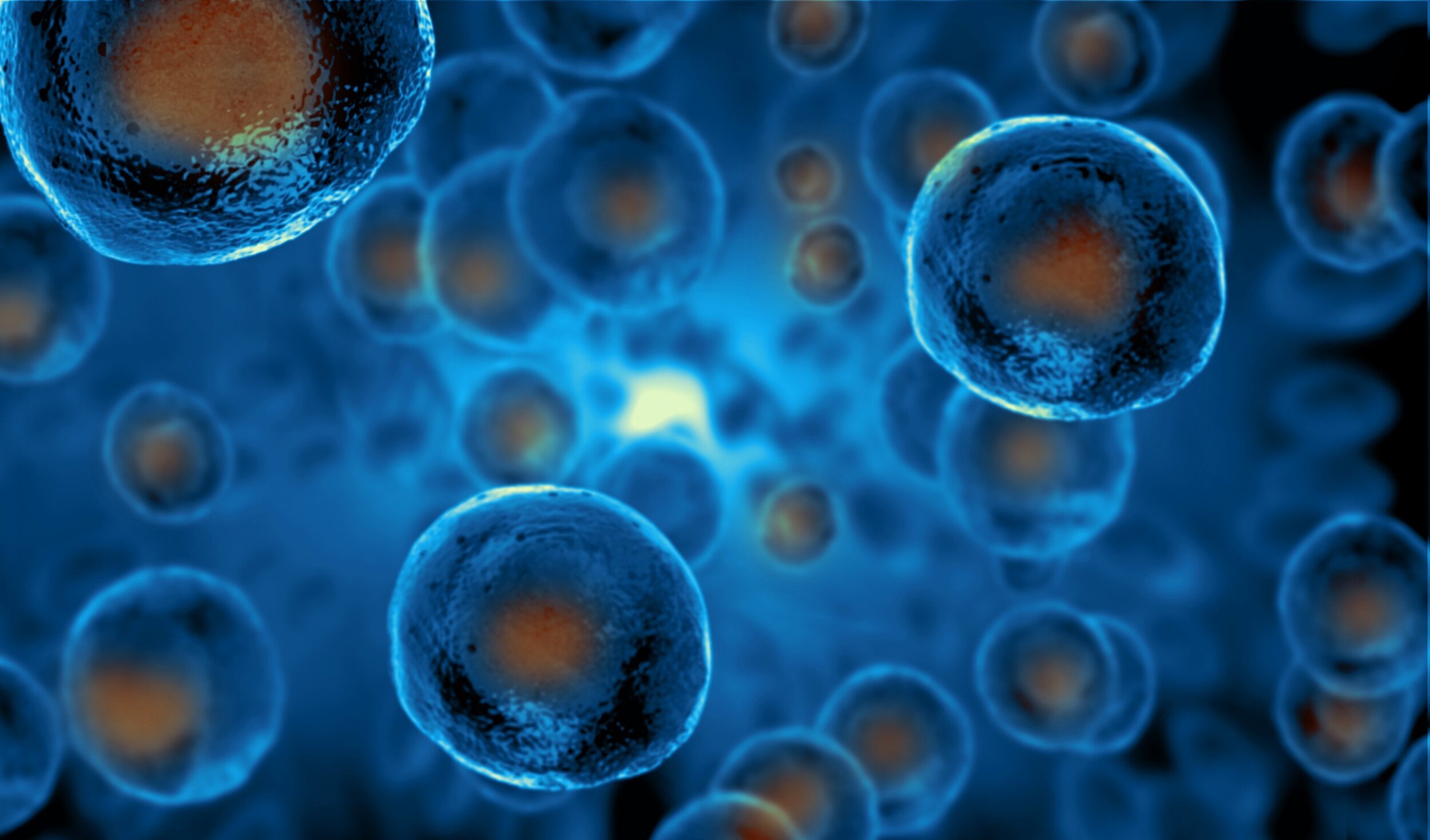
Stem cells are administered either surgically, or via injection into the arthritic joint. Much like the image above, your orthopaedic surgeon will utilize medical imaging in order to identify where on the body, the stem cells are to be administered. Depending on the severity of the injury, either of the two options will be applied. When the stem cells are being administered, your orthopaedic surgeon will more than likely utilize mesenchymal stem cells that have been harvested from the individual's body. 
 

Studies have identified that joint-resident MSC’s are integral to the facilitation of joint regeneration stability, and also regulate cell turn over and tissue repair. A regenerative microenvironment is the outcome of introduction of MSCs into an arthritic joint. The reversal of OA is now possible with these newly understood mechanisms. 

Cartilage Degeneration 

With an understanding of OA biological processes that trigger OA, the primary aim now is to identify the means to minimise cartilage degeneration. Resident joint MSCs work to modulate local inflammatory and immunologic responses. Thus far, MSCs seem promising for both early stage OA all the way to more advanced joint degeneration. 

RESTORATIVE SITES IN OA

The knee joint forms a joint capsule where all the bones meet in the hinge mechanism. Within the joint is a fibrous membrane that is composed of connective fibrous tissue. The nervous system permeates this area introducing feeling, while muscles and tendons facilitate the motion and flexibility of the joint. The synovial membrane in the joint, is the point where the special mesenchymal tissue resides. This is the point of regeneration. As a regenerative site, there is a dense net of capillary vessels that provide the nutrients that are distributed via processes such as diffusion. All joints have a fibrous membrane structure that varies in thickness in different parts of the body. For this reason, it is possible to transfer some of the MSCs from the fatty parts of the body into sites that need to be regenerated. They can’t always be generated in situ, but they can be transferred from a pre-existing site.

Within the synovial membrane complex is the important function of generating synovial fluid, as well as the sustenance of the Extracellular Matrix that is a cell host. Synovial fluid is a key element in the health of the cartilage. It supports the superficial and middle zones of the cartilage, and provides them with nutrients. 

The General Properties of MSCs 

MSCs are members of the stem cell family. The innate ability of all stem cells is to generate mesenchymal lineages such as fibroblasts, chondroblasts, adipocytes, muscle-cells, osteoblasts and other mesenchymal lineages [1]. This ability of the stem cell to forge differing cell groups is what facilitates the in vivo proliferation and differentiation in the regenerative microenvironment that is the joint. Their mechanisms of action include autocrine and paracrine effects. Medical research is still delving into the various mechanisms of action of the MSC cells. They have various interactions with critical receptors in the joint capsule. One thing that activates the already resident MSCs in the joint is presence of tissue injury and inflammation. The regenerative mechanisms are activated once these conditions are induced. Local immune modulation and trophic effects are two of the primary functions of the MSCs. MSCs as a heterogenous stem cell population that generate the relevant cells needed for healing in a tissue-specific microenvironment. They adapt to their environment. 

MSC Exosome Research 

In the past decade, research in MSC has resulted in several experimental models that have ultimately led to clinical applications. Since the MSC’s show great promise in promotion of healing and tissue regeneration there is a lot of interest in their utilization. What is the current concern of researchers is to ensure that the survival rate of MSCS during and after transplants is understood. Survival rate verification is currently to be determined, and future research is still to understand the full mechanisms of action once the transplanted cells are placed in the host tissue. Unlike drugs whose concentrations and paths are traceable, there is still a lot to be learned from the Stem Cell research. Sometimes in the research process, when MSCs were intended for proliferation they would act in an immune-regenerative function instead. This inability to specify the mechanism is what is limiting the use of the MSCs  and also the progress of usage in therapies. The root of this condition is linked to a process known as paracrine action. Studies have linked this outcome to the secretion of exosomes. These membrane-enclosed vesicles favor regeneration. Knowing how to adjust the lever to the right specificity will be the key to a successful future. 

Reference Article: 

  1. ScienceDirect: https://www.sciencedirect.com/science/article/pii/S2352320420300651

Gordon+Slater+Logo.jpg

What You Need To Know About Equinus

Academy Foot and Orthotic Clinics.jpg

Image Credit: Academy Foot And Orthotic Clinics 

Authors: Gordon Slater| Tandose Sambo

“The higher your energy level, the more efficient your body. The more efficient your body, the better you feel and the more you will use your talent to produce outstanding results.”-Tony Robbins

What Is Equinus?  

Equinus is a limitation of the upward bending motion of the ankle joint. While most persons can create somewhat of a right angle between their feet and their lower legs, individuals with equinus are unable to do this. As a condition that affects the feet, equinus can occur in either one or both of the feet. If both feet are affected, the severity is different in each foot, as the body adjusts in order to manage the body’s weight. 

If left unmanaged, individuals with equinus often find ways to compensate for their limited ankle mobility. Without adequate orthopaedic care, the body will develop additional foot, leg, or back problems. Many persons, for instance, place the majority of their weight on the ball of the foot.

What Causes Equinus?

Equinus has several identified root causes. The first root cause can be attributed to a tightness in the Achilles tendon, or calf muscles. The restriction can be induced either as an inherited trait, or developed during later stages of life. Additional causes of the onset of equinus includes limited mobility of the leg while it’s in a cast, during the process of wearing crutches, or even with the wearing of high heeled shoes. High heels are shoes that women must be careful in wearing because they cause several different foot ailments including hammertoes. Diabetics are also another group that is prone to developing Equinus. During the diabetic process, the condition can affect the Achilles Tendon, and ultimately cause a tightening of the tendon. Underlying neurologic disorders, also associate with diabetes are also responsible for spasms that can affect the ankle joint.

For an externally induced incident of Equinus, if an individual has a fracture of the leg, a fragment of bone can dislodge, and cause a restriction in motion of the ankle. With a variety of root causes that can cause the condition, it is important for the appropriate treatment plan to be put in place that will facilitate restoration of the feet.

Foot Problems Related to Equinus

Depending on how a patient compensates for the inability to bend properly at the ankle, a variety of foot conditions can develop, including[1]:

  • Plantar fasciitis (arch/heel pain)

  • Calf cramping

  • Tendonitis (inflammation in the Achilles tendon)

  • Metatarsalgia (pain and/or callusing on the ball of the foot)

  • Flatfoot

  • Arthritis of the midfoot (middle area of the foot)

  • Pressure sores on the ball of the foot or the arch

  • Bunions and hammertoes

  • Ankle pain

  • Shin splints 

Diagnosis of Equinus

The best way to classify a case of Equinus, is to consult with an Orthopaedic Specialist. From a medical perspective, equinus is often diagnosed, when the patient is unable to achieve 10° of ankle joint dorsiflexion[2]. What patients often experience before the severe cases of Equinus are the symptoms that are associated with the condition. 

There will be a variety of tests that are undergone, in order to diagnose if a patient has equinus. Your foot and ankle surgeon will initially test the range of motion of the ankle. This is done in two states: when the knee is flexed (bent) or straightened. Range of motion tests will identify any tightness in the muscles or tendons. 

For an internal view of the ankle, X-rays will also be done. From the aforementioned introduction, there may be some neurologic tests that will be done, in order to ensure that the nervous system is operating as it should. 

Nonsurgical Treatment of Equinus

Surgical treatment of any orthopaedic condition, is often an action of last resort. In the initial stages of Equinus, there are a series of actions that your orthopaedic surgeon will prescribe in order to keep your Equinus under control. These include[1]:

  • Night splint. The foot may be placed in a splint at night to keep it in a position that helps reduce tightness of the calf muscle.

  • Heel lifts. Placing heel lifts inside the shoes or wearing shoes with a moderate heel takes stress off the Achilles tendon when walking and may reduce symptoms.

  • Arch supports or Orthotic devices. Custom orthotic devices that fit into the shoe are often prescribed to keep weight distributed properly and to help control muscle/tendon imbalance.

  • Physical therapy. To help remedy muscle tightness, exercises that stretch the calf muscle(s) are recommended.

From medical research: “Treatment for equinus should be aimed at increasing ankle joint dorsiflexion to facilitate normal gait mechanics. In most cases, nonspastic forms of equinus tend to be more treatable and easily corrected than the spastic neuro-induced forms of equinus. A variety of conservative measures can be employed, including stretching, bracing, and orthotic devices.

The merits of manual stretching for the gastrocnemius muscle have been a topic of debate, but studies have demonstrated that favorable results are possible when stretching is done correctly even for short time periods. Grady and Saxena found that manual stretching for five minutes per day over six months increased dorsiflexion by an average of 2.7°.23,33 Similarly, Macklin and colleagues were able to elicit excellent results in a group of runners with equinus contractures. Their results produced an average increase in ankle joint dorsiflexion from 5° to 16° following an eight-week stretching program.”[2] 

When Is Surgery Needed for Equinus?

If there is an instance of severe Equinus, surgery may be the only option that will ensure an adequate recovery. If there is a bone fragment or a tight tendon, it will be important for surgery to be done. As an inherited trait, the calf muscle tightness is one that often manifests later in life, and surgical treatments can help to alleviate this condition. The surgical path that is decided by your orthopaedic surgeon will be dependent on the case. There will be a series of decision matrices involved, such as the utilization of the Silfverskiöld test to identify your Equinus classification. Depending on the classification, the individual's surgery will involve either a gastrocnemius recession or an alternate surgical path. With this procedure, there is a noted increase in ankle joint dorsiflexion (range of rotation) of 18.3°. 

With such a positive gain to be achieved from surgery, persons with equinus can feel comforted that they will be able to achieve an improvement in their mobility with either non-surgical or surgical procedures.

References: 

  1. FootHealthFacts: https://www.foothealthfacts.org/conditions/equinus

  2. LER: https://lermagazine.com/article/equinus-its-surprising-role-in-foot-pathologies

Gordon+Slater+Logo.jpg

Haglund's Deformity

haglund-s-deformity-is-a-bump-that-appears-on-the-back-of-the-heel.jpg

Image Credit: Medical News Today 

Authors: Gordon Slater |Tandose Sambo

“The human body has been designed to resist an infinite number of changes and attacks brought about by its environment. The secret of good health lies in successful adjustment to changing stresses on the body.” – Harry J. Johnson

What Is Haglund’s Deformity?

When a large, bony protrusion develops on the back of the heel, this condition is known as Haglund’s deformity. As a bony enlargement, the condition causes the soft tissue in the surrounding area, near to the Achilles tendon to become irritated during daily activities, such as the wearing of shoes. When the heel area becomes irritated, conditions such as bursitis can develop. With a variety of treatments both non-surgical and surgical, it is possible to treat this condition, and restore normal life activities. 

Causes

With a variety of root causes for this condition, one of the most frequent causes of Haglund’s deformity is actually induced via the wearing of pump style shoes that can aggravate the tissues and the bones in the heels. It is therefore important to wear the right shoes: 

What is the right shoe? 

The right shoe provides the feet with a mixture of cushion, support and comfort. The anatomy of the foot is such that it supports the body on a hinge joint. The appropriate alignment of the feet is critical to so much, inclusive of the health of the spine. The right fit of the shoe should be such that it fits snugly, and there is at least 1-1.5 cm of space at the end of shoe. A few key characteristics of shoes that should be factored in include: 

  1. A stiff back for appropriate heel support 

  2. Flexibility to facilitate the motion of your feet

  3. Appropriate arch support - The arch of the foot is part of its engineering design that facilitates its ability to support the body appropriately. Shoes will not all be perfectly designed, so with the appropriate inserts, shoes can be customized to facilitate that additional comfort. 

  4. Heel height - Heel height is very important for the support of the feet and the body. The body was designed for an optimum height. The higher an individual is off the ground, the higher will be the center of gravity of that individual. Any object with a high center of gravity will have the tendency to fall, compared to an object with a lower center of gravity. Women are aware of the fact that with higher heels, they do feel the instability that comes with being elevated. It will be important for an individual to ensure that they match their heel height with their personal height. 

Because the wearing of shoes such as pumps can develop Haglund’s Deformity, the condition is often known as “pump bump”. The rigid backs of the heels, coupled with the already enlarged heel, will induce the generation of the growth. For those in the sporting realm, ice skates can also induce Halgund’s Deformity. From a genetic perspective, if you have the following noted characteristics:

  • A high-arched foot

  • A tight Achilles tendon

  • A tendency to walk on the outside of the heel,

you will likely develop Haglund’s Deformity. 

Symptoms

  • A noticeable bump on the back of the heel

  • Pain in the area where the Achilles tendon attaches to the heel

  • Swelling in the back of the heel

  • Redness near the inflamed tissue

Source: Reference[1] 

Diagnosis

During a consultation with your Orthopaedic surgeon, you will then be presented with a suitable course of action. There will be more than one foot condition that can lead to an enlargement of the heel area, so carefully ensure that you are thoroughly assessed. In some instances, it is highly likely that you may have a condition such as Achilles Tendonitis, instead of Haglund’s deformity. Medical tests will be the most efficient way to determine what the diagnosis of your condition will be. 

Via the utilization of equipment such as an X-ray, the internal conditions of your heel will be visible to your doctor. He can then take the time to actually determine your condition. The prominent heel bone will be visible from the lab results. Once the diagnosis is indeed Haglund’s Deformity, your doctor can also take the necessary steps to ensure that you’re taken care of. This includes the manufacture of your custom orthotic device, a foot insert, that will help to relieve your heel pain. 

Nonsurgical Treatment

In the instance where your condition is in its early stages, you may not need surgery in order to treat your bursitis. What works well, is the utilization of various treatments and medications that will work for management of your condition. These treatments include: 

  • Medication. Oral nonsteroidal anti-inflammatory drugs (NSAIDs), such as ibuprofen, may be recommended to reduce the pain and inflammation. Ice. To reduce swelling, apply an ice pack to the inflamed area, placing a thin towel between the ice and the skin. Use ice for 20 minutes and then wait at least 40 minutes before icing again.

  • Exercises. Stretching exercises help relieve tension from the Achilles tendon. These exercises are especially important for the patient who has a tight heel cord.

  • Heel lifts. Patients with high arches may find that heel lifts placed inside the shoe decrease the pressure on the heel.

  • Heel pads. Pads placed inside the shoe cushion the heel and may help reduce irritation when walking.

  • Shoe modification. Backless or soft backed shoes help avoid or minimize irritation.

  • Physical therapy. Physical therapy modalities, such as ultrasound, can help to reduce inflammation.

  • Orthotic devices. Custom arch supports control the motion in the foot.

  • Immobilization. In some cases, casting may be necessary.

Reference[1] 

When Is Surgery Needed?

Surgery is always the treatment of last resort, because it is so invasive in nature. From the list of non-surgical methods outlined above, a combination of the treatments works well in order to achieve a comfortable standard of living. In those instances where nonsurgical treatment fails to provide adequate pain relief, surgery may be needed. Your orthopaedic surgeon will then prepare an adequate plan for your to ensure that your Haglund’s Deformity protrusion is removed. At best, wear your orthotic devices and keep your feet as comfortable as possible, until your surgery is done.

Post-operative management

With all treatments, there’s a time line for recovery.  Full rehabilitation after Haglund's resection can take up to one year. Here is a solid plan for healing[3]:

Day 0 - 10

  • Oedema management:

    • Rest - Active rest

    • Ice

    • Elevation - for first 10 days of recovery period

    • Compression

  • Mobilization:

    • Patients are mostly put in moon boot or below-knee backslab

    • Non-weight bearing mobilization: 3 days to 2 weeks (as per surgeon)

  • Lower leg strengthening exercises

  • Achilles stretches (contra-indicated if Achilles was injured or if it was detached and re-attached intra-operatively)

  • Return to full activity at 6 weeks post-surgery.

Day 10 - 6 weeks

  • Mobilization:

    • Resection without Achilles debridement/repair: Moonboot with raised heel

      • Progress from toe-touch weight bearing to partial weight bearing to assisted full weight bearing in the 4 week period

    • Resection with Achilles debridement/repair: Moonboot or cast in equinis

      • Non-weight bearing 4 weeks

  • Oedema management as needed

6 - 12 weeks

  • Mobilization:

    • Resection without Achilles debridement/repair:

      • Wean moonboot wear

      • Full weight bearing

  • Resection with Achilles debridement/repair: Moonboot in equinis

    • Progress to moonboot is cast was initially required

    • Progress weight bearing from toe-touch weight bearing to partial weight bearing to assisted full weight bearing over 4 weeks

  • Oedema management

  • Start scar management as soon as wounds are fully healed

Week 12+

  • Mobilization: Wean off moonboot and mobility assistive devices if still in use

  • Exercises:

    • Strengthening and endurance of foot and ankle muscles

  • Oedema management

References: 

  1. FootHealthFacts: https://www.foothealthfacts.org/conditions/haglund%E2%80%99s-deformity

  2. HealthLine: https://www.healthline.com/health/haglund-deformity#TOC_TITLE_HDR_1

  3. Physiopedia: https://www.physio-pedia.com/Haglund%27s_deformity

Gordon+Slater+Logo.jpg

Diabetic Peripheral Neuropathy

1800x1200_what_to_do_about_diabetic_peripheral_neuropathy_slideshow.jpg

Image Credit: WebMD 

“Health is a large word. It embraces not the body only, but the mind and spirit as well; …and not today’s pain or pleasure alone, but the whole being and outlook of a man.”- James H. West

What Is Diabetic Peripheral Neuropathy?  

Diabetes is one of the most common health conditions that afflicts the population. As a systematic ailment, there are often downstream implications of the disease that are experienced by patients, if they don’t take the necessary precaution to control their diabetes. One such condition is diabetic peripheral neuropathy - a nerve damage induction that can be potentially serious if not managed. As the nervous system is compromised, the patients with peripheral neuropathy will experience symptoms such as loss of feeling in their extremities (arms and legs) and because they can’t feel, they can often develop cuts or ulcers on the skin as they go about their daily activities. The parts of the body affected by diabetic neuropathy are often the arms, hands, legs and feet.  Statistically, up to 70% of all diabetics eventually do develop peripheral neuropathy. The extremes of the condition are dependent on the patient and their lifestyle.

All health conditions can be managed with the appropriate internal chemistry. Medical research has indicated that diabetics who manage their blood sugar levels well, can avoid or minimise the odds of even developing peripheral neuropathy. Via the right diet, the peak states of health will be achieved. 

Three different groups of nerves can be affected by diabetic neuropathy[1]:

  • Sensory nerves, which enable people to feel pain, temperature and other sensations

  • Motor nerves, which control the muscles and give them their strength and tone

  • Autonomic nerves, which allow the body to perform certain involuntary functions, such as sweating 

With all systematic conditions, there is always a delay between the onset and the full development of peripheral neuropathy. In some instances, there are some patients who have had peripheral neuropathy before they even had diabetes. The fusion of the two conditions is what patients need to be careful of.  

What causes Diabetic Neuropathy?

With diabetic neuropathy, there is a direct correlation between the extent of diabetic neuropathy and the management of critical diabetic parameters such as blood sugar levels. Although there’s no 100% guarantee that you won’t develop diabetic peripheral neuropathy if you stay in a good health care regime, the odds will significantly be higher, and you will achieve your peak longevity as a diabetic patient. With the onset of high blood sugar levels, the blood vessel impact from the internal chemistry is what will affect the nervous system.

Anticipated symptoms of Diabetic Neuropathy include[1]:

  • Deformities (such as bunions or hammertoes) resulting from motor neuropathy may cause shoes to rub against toes, creating a sore. The numbness caused by sensory neuropathy can make the patient unaware that this is happening.

  • Because of numbness, a patient may not realize that s/he has stepped on a small object and cut the skin.

  • Cracked skin caused by autonomic neuropathy, combined with sensory neuropathy’s numbness and problems associated with motor neuropathy, can lead to developing a sore.

Symptoms of Diabetic Neuropathy[1]:

For sensory neuropathy:

  • Numbness or tingling in the feet

  • Pain or discomfort in the feet or legs, including prickly, sharp pain or burning feet 

For motor neuropathy:

  • Muscle weakness and loss of muscle tone in the feet and lower legs

  • Loss of balance

  • Changes in foot shape that can lead to areas of increased pressure 

For autonomic neuropathy:

  • Dry feet

  • Cracked skin

With appropriate discussions, your orthopaedic surgeon will be able to determine if you have diabetic peripheral neuropathy. As an anticipated condition, there will be routine practices that will be incorporated into your healthcare routine that will indicate if you’re developing this condition, or are stable. The first stages of treatment will involve the management and control of the patient's blood sugar level. Based on the aforementioned correlation, it will be important to monitor this parameter for an indication of internal health. Medications for pain are prescribed according to the symptoms that the patients are experiencing. Physical exercise, and consultation with individuals such as physiotherapists, will facilitate the management of body circulation. 

DAILY FOOT INSPECTION

As a start, you can consult with your orthopaedic surgeon to identify what items will be beneficial to your at home foot care kit. This is a great option, if you’re diabetic, and need to inspect your feet regularly, preferably on a daily basis. Instead of foot inspection feeling like a chore, it will feel like a luxury and you’ll want to do it. If you can, as a diabetic, indulge in the following:

  1. Foot Spa: With a wide range of options from the super affordable to the super luxe, it is possible to purchase for yourself an at home foot massager. With this massager, you can facilitate the massaging of your feet, and improve the circulation through your entire feet and ankles. The soothing warm water will relax both your feet and your mind. Interestingly, if you really want to go low budget, you can add some dish washing liquid to your water. The agent is antimicrobial and moisturizing. Additionally, you can purchase foot soaks from your local pharmacy or beauty supply store. Jazz up the experience with a little lavender soak, and you’ll be healing and relaxing at the same time. 

  2. Epsom Salts: The versatility of Epsom Salts cannot be stated enough. From an internal perspective, it is the perfect body detox for your liver. If you ever feel your energy levels on a low, it may be that your body is toxic. Toxicity is induced by the ingestion of preservatives with time. You can either detox via internal ingestion of the salts, or you can detox via the osmosis process. In your foot spa, or a basin, you can create a salt solution. Fill the bath just about half way, and add half a cup of Epsom salts. If you have any blisters, they will heal with time. Additionally, via osmosis, with a concentrated salt solution, you can detox as the salts in the water will pull toxins from your body through the pores in the soles of your feet. The soles of your feet are actually very sensitive and porus.  

  3. Foot Scrub: After you have soaked your feet, you can facilitate exfoliation by using a foot scrub to massage the feet. You can create your own scrub via a mixture of coffee (after you’ve percolated it and have the ground coffee left over), coconut oil and brown sugar. Apply gently, and let the scrub nourish your feet. 

  4. Foot Cream: With aromatherapy becoming more mainstream, you can actually determine what scents to apply to your feet in order to give yourself a boost. If you’re heading to sleep you can apply a lavender lotion. These are very inexpensive and you can find them in your local pharmacy, near to lotions and candles. If you want to energize your body, you can actually apply an invigorating scent like a mint or citrus scent that will energize the senses once they are stimulated.

Once you are finished, you can simply go through the process of either placing your feet into socks, or if you’re heading out for the day into your favorite comfortable sandals. 

With all health concerns, prevention and maintenance is better than cure. Take the time to actually investigate the conditions that induce diabetic neuropathy. Where possible, with an appropriate health care plan, you will achieve your peak health. 

References: 

  1. Diabetic Peripheral Neuropathy: https://www.foothealthfacts.org/conditions/diabetic-peripheral-neuropathy

  2. Diabetic Peripheral Neuropathy: https://www.webmd.com/diabetes/peripheral-neuropathy-risk-factors-symptoms

Gordon+Slater+Logo.jpg

Articular Cartilage Regeneration - Advances in Medical Growth Factor Generation

worn-knee-cartilage.jpeg

Image Credits: Sports Health 

Authors: Gordon Slater | Tandose Sambo

“Health is a large word. It embraces not the body only, but the mind and spirit as well; …and not today’s pain or pleasure alone, but the whole being and outlook of a man.”- James H. West

Introduction

Progress is a continuous effort. In all fields, there’s a desire for growth and change. One of the best things that has happened in 2020 is the advancement of aerospace and the resuming of activities that are gearing us towards the limits of our solar system. There is a Universe within us as well, and there are always continuing increases in the technologies that are dedicated to improving our health and well being. In the area of cellular and molecular biotechnology, there is a noticeable transition toward the over-production of therapeutic and anabolic growth factors. With this development, it will one day be a useful application for the treatment of osteoarthritis (OA). As a regenerative treatment, the overproduction of growth factors will facilitate self healing, once the relevant environment is created. 

The vision for such therapies is the utilization of GP293 cells to generate the therapeutic proteins and anabolic growth factors that will enhance the restoration and regeneration of the cartilage. Scientific studies have indicated that there is a reduction in healing capacity with the onset of the aging process. With growth factor enhancers however, this will no longer be a limitation. Utilizing the Tissue Gene-C therapy, biologic treatments will be facilitated in this regard. With live cells as a baseline, the ability for utilization of cells to be biological drug delivery systems will be the wave of the future. 

With new innovations currently being developed and deployed, regenerative medicine is emerging as the go to therapy for degenerative diseases such as OA. As a start, as a patient, you can imagine that an injection will one day...be able to restore your joints.

What are Growth Factors? 

Growth Factors (GF’s) are proteins that facilitate the internal healing processes. The primary function of growth factors is to catalyze growth, proliferation, migration, survival and differentiation of target cell types. In the physical realms, catalysts are elements in a reaction that speed up a reaction. Biological catalysts like growth factors facilitate similar mechanisms in biological reactions. Their presence helps to improve reaction rates, and generation rates of critical healing components in your body. Growth factors are regulators and determine what the specialised function and phenotype of cells will be, as well as how they will interact with other elements in the cells in the genetic expression process. GF’s have been found to be effective on the majority of cell types, with the exception of cells like mature neurons. 

As biological catalysts, the GF’s are small and stable polypeptides which are naturally secreted within the body’s cellular system. Secreted into the extracellular matrix, they then get to work and regulate several body functions. It’s really an amazing biologic marvel. When sourced from stem cells, the GF’s generated can facilitate the healing process and stabilize tissue regeneration and maintenance, as well as heal wounds in the skin. Articular cartilage benefits greatly from the presence of stem cell derived GF’s. Studies have shown that in terms of the articular cartilage classification both IGF-1 and FGF have proven to be beneficial to articular cartilage repair. Within the transforming growth factor-β (TGF-β) superfamily, the system is encoded by 33 genes that work to facilitate wound healing and immunoregulation. 

TGF-β has action on articular cartilage, that is found to be dependent on a variety of factors. The current studies indicate that in vivo and in vitro performance can often differ. Some of the impactors to healing can include the nature of the internal environment of the patient. It reacts differently for instance in a young healthy joint, versus an ageing joint. Studies have shown that TGF-β is actually more active in OA joints. This indication is one of the reasons that it is being targeted as a treatment of OA. As a smart reactor, the TGF-β is showing signs of specificity, which is what is needed for any drug - be it synthetically generated, or biologically induced. The lock and key and target mechanisms are key to ensuring that specific conditions such as OA are treatable, without any adverse side effects to the body. 

How are these Growth Factors delivered? 

With the current state of the art technology in cellular and molecular biotechnology, live carriers of the GF will be injected into the cavity of the knee joint. Via the utilization of what is classified as “transfected and irradiated protein packaging eukaryotic cell lines”, these carrier cells will be the generators of the GF proteins. Generating the relevant cells for healing, the appropriate combinations of the GF’s necessary to heal the joint will be easily delivered. The healing cocktail which typically will contain cells such as chondrocytes and mesenchymal stem cells (MSCs) to enable maximum success. 

What is Osteoarthritis? 

Statistically, osteoarthritis is the most prevalent joint condition. Induced by a multitude of root causes, it is ultimately one of those conditions that has a wealth of treatments ranging from the utilization of wholistic healing methods such as joint healing oils, or the administration of surgery to facilitate the healing or replacement of the joint. 

Joints are the union of two or more bones. As a mechanical unit, the free flow of the joint is facilitated by the natural lubricating agents known as cartilage. Cartilage is soft protective tissue that lines the exterior of the bone. Under the condition of osteoarthritis, the cartilage degenerates and exposes the bones to each other. The painful condition results in limited mobility of the patients. While the majority of osteoarthritis cases take place in older patients, who have reduced healing capabilities, athletes and active individuals who place stress on their joints, can also develop arthritis. If left untreated, arthritis can induce disability. 

Causes of Osteoarthritis

It was highlighted in the previous introduction that cartilage degeneration is what ultimately leads to the joint damage associated with osteoarthritis. Joint damage as a whole has several root causes. If you had an injury for instance that resulted in the dislocation of a joint, there can also be resultant joint damage that can also inflict symptoms of osteoarthritis. Osteoarthritis does have genetic links, so ensure that your family medical history is assessed, and you’ll be able to identify with your doctor’s help, if you or your family members are at risk of the condition.

The Science of Cartilage 

As a tough, flexible coating to the bone, when cartilage degenerates, the friction between the joint’s bones can cause the exposed surfaces to become jagged. Cartilage, with time and age, is not a naturally self healing mechanism, because there is limited blood supply to the area. Biologics however, is beginning to enable the facilitation of healing by introducing the appropriate growth factors that will enable the cartilage to rebuild. The symptoms that patients experience include: pain as an initial indicator of a joint issue. With inflammation, the joint will swell and tenderness be experienced. As the joints lose their flexibility, there will be stiffness experienced, often in the morning upon rising. Your orthopaedic surgeon will assess your condition and determine the best way for you to approach your treatment. With all health conditions, with time, you can actually initiate healing via the facilitation of the right internal chemistry. A good and proper diet, can reduce the internal inflammation that you experience. 

THE STAGES OF OSTEOARTHRITIS

Osteoarthritis has the following five classifications:

Stage 0 - Normal 

Stage 1 - Minor 

Stage 2 - Mild 

Stage 3 - Moderate 

Stage 4- Severe

Consult with your orthopaedic surgeon to identify what stage of the progression you are on, if you are diagnosed with osteoarthritis. Cartilage loss is most prevalent in the higher tiers of the osteoarthritis scale. You are also at risk of increases in swelling and inflammation, and joint instability. 

ORTHOPAEDIC CONSULTATION

There are some conditions that take a long time to actually develop and diagnose. Osteoarthritis is one of those conditions. It is often during the periods of intense pain that one would be led to an orthopaedic specialist for the root cause of this discomfort. During your consultation, your orthopaedic surgeon will usually utilize a diagnostic tool such as an X-ray or an MRI image, in order to see the internal functions of the joint. For further details of your internal chemistry, blood tests, joint fluid tests and other tests can be conducted in order to eliminate additional root causes. From the surface, Rheumatoid Arthritis and Osteoarthritis do have almost identical symptoms. The critical tests will provide your orthopaedic specialist with the decision making tools that will enable your condition to be diagnosed. As a progressive and degenerative condition that causes the joints to stiffen, mobility is often a challenge with OA. 

How Growth Factors can improve OA

Based on the aforementioned mechanism of generation, GF’s are proving to important to generation and synthesis of articular cartilage. In the orthopaedic realm, bioactive GFs are currently being trialled in order to test their ability to heal chrondal injuries and inhibit the arthritic process. Scientific studies have identified a few relevant growth factors, that have been outlined below: 

Types of Growth Factors.PNG

With the advent of the application of a combination of these growth factors, it will be possible to facilitate the healing of cartilage, as well as bone repair and regeneration. As the demand for recombinant therapeutic proteins is on the rise, the accumulated knowledge and applications of these therapies will one day help to combat a condition that plagues many. With the current potential list of growth factors, these novel biological agents for cartilage regeneration show a very promising future. As study and treatments evolve, the cartilage degeneration that is identified in the age group of individuals over 50, it will be possible to “turn back the hands of time” and restore the limbs and joints to full capacity. Additional GF therapies will have applications in areas such as sports injury. 

For further details, consult with your orthopaedic specialist. You may actually qualify for a trial if the conditions and research facilities are in your local area. 

Reference Article: https://www.preprints.org/manuscript/202007.0674/v1/download


Gordon+Slater+Logo.jpg

Orthopaedic Gene Therapy - Implications for Future Research

Inquiries Journal.jpg

Image Credits: Inquiries Journal 

How does Gene Therapy work?

With the advent of therapies within the biologic realm, the focus of medicine now and in the future is to actually facilitate internal healing of the body. With increasing studies of how healing mechanisms are carried out in the body, it will be possible for the appropriate levers to be pulled, that will facilitate the enhancement, or even acceleration of the healing process in the human body. 

One of the focus areas of internal healing is via the utilization of technologies such as gene therapy. Designed to introduce genetic materials into cells, as a means to compensate for any abnormalities in the genes, or to ensure that beneficial genes are being manufactured by an organism, gene therapy is really high level engineering that will facilitate future biological changes within a living system. 

Genes are the underlying foundation of our human existence. Understanding their mechanisms will be the key to ensuring that the appropriate functional proteins are being generated by a system. If a gene with an appropriate carrier vector is delivered into a cell, it will be able to carry out its necessary functions. The utilization of  modified viruses is one such way that a new gene can be delivered into a cell. Alternative methods also include the introduction of the genes to some extracted cells from a patient, in a laboratory. Once the genes have been introduced to the cells, after the appropriate incubation period, they will be returned to the patient. 

While gene therapy is still in its infancy, there is still lots to be learned in order to ensure that the full mechanisms and applications of the therapy can be applied to the treatment of cases. Within the orthopaedic realm, there is ongoing research being carried out, to ensure that the applications of gene therapy can be facilitated.

Orthopaedic Gene Therapy 

Orthopaedic Gene Therapy is an area of study that has spanned a few decades. Scientific studies have indicated that with the previous work that was done, there have been successful preclinical trials, and as a result the future for orthopaedic applications of genetic therapies looks very promising. At this point in time, many therapies that are under study are in the process of achieving FDA approval. Because of the underlying uncertainties that are associated with gene therapy, the track to FDA approval is still being worked on. There is still a lot to be learned about gene therapy, and as a result, the FDA often views the research as unsafe and risky. For projects that are deemed as having little ROI, the pharmaceutical industry will not be inclined to fund such activities. 

Of course, the beauty of science and technology is that it is possible to self fund activities. Many start up companies have decided to challenge themselves to actually do the research that will enable the field to progress. Being bold enough to take on such critical challenges in the medical realm will only reap great rewards for those who are able to master the healing mechanisms. 

The successful accomplishment of gene therapy will require the close interaction of scientists, clinicians and the regulatory bodies who will facilitate the approval and distribution to the masses. Science and technology is a realm that takes decades for success. With a little focus, perseverance and determination however, there will be a possibility for success. This has been revealed and documented in all realms of the scientific sphere. In the aerospace industry for instance, many of the rockets that are currently being deployed, were under construction for years before they were finally fit for orbit. The medical realm shouldn't expect itself to be any different. There will be constraints, but nothing that will be impossible in the long run. Gene therapy is a very promising area of research, and with a sense of optimism, the orthopaedic realm will definitely benefit from the upsurge in the field. 

How does Gene Therapy Work? 

Via the ability to modify genes, it will be possible for mutations to be eliminated within a living organism. With the utilization of vectors, gene transfer will allow the therapeutic genes to enter a living system. Once in the living cell, the appropriate level and duration of transgene expression will be facilitated. Scientific studies have now facilitated the improvement in the knowledge of the level and duration of transgene expression. Via the utilization of biological promoters and other regulatory mechanisms, it will be possible to achieve the desired outcome. 

What Orthopaedic conditions are treatable by Gene Therapy? 

Four main areas of orthopaedic research have been found to be useful for the application of Gene Therapy in the orthopaedic realm. These four areas include: 

Mendelian disorders 

Tumors 

Arthritis and other Joint Diseases 

Tissue Repair and Regenerative Medicine 

Additional Genetic Diseases 

Arthritis is one of those areas that is statistically highest in the orthopaedic realm. With the ability to directly inject the critical gene modifiers into the diseased joints, it is possible to monitor any improvements in the healing process. Arthritis is a joint ailment, and the joint cavity does provide the space to inject healing solutions such as gene modifiers and even stem cells. The mechanism of the joint, with the synovium a joint component that allows gene delivery, the gene vectors will engage with the synovial cells and the interaction will start the gene modification process. 

Once the synovial cells are genetically modified, the synovial cells can then start a new expression, and deliver the appropriate transgene products into the joint. The enhanced healing of the arthritic joint can then be facilitated, and the joint will heal itself.

With arthritis being such a prevalent condition, improving the health of patients with this condition will have great advantages in the medical realm.  

Tissue Engineering and Regenerative Medicine 

Another statistically high orthopaedic occurrence is that of the sports injury. During sports injuries, various soft tissues are impacted in a patient, and their healing time may range depending on the material. While tissues like cartilage can repair relatively fast, there are other hard materials such as bone that takes a longer time to repair, and often requires some scaffolding to hold the joint in place while the system is regenerating itself. 

Orthopaedic gene therapy is a technology that can benefit the patient. With an improved understanding of the repair processes that are involved in healing a joint, it will be possible to start the healing process either in the laboratory or in vivo. Current studies have identified the appropriate carriers for healing agents of tissue and bone, and the appropriate healing mechanisms are being studied and trialled. There is always a pressing need to heal bone, and soft tissues such as tendons and ligaments. With the appropriate clinical trials, anticipation of treatment to patients will one day be available. 

In conclusion, the orthopaedic realm is still a place with lots of potential to grow and develop. Once successful, the treatment of conditions such as arthritis and bone healing will be facilitated via this method. With orthobiologics, the sky is still the limit. Science and technology is always an area that is growing and evolving. Talk to your orthopaedic surgeon about treatments today that you can trial. 

References: 

  1. Orthopaedic Gene Therapy: https://www.ncbi.nlm.nih.gov/pmc/articles/PMC3264489/

Gordon+Slater+Logo.jpg

Deep Vein Thrombosis (DVT) - Causes and Treatments

Awareness Days.jpeg

Image Credits: Awareness Days 

“Health is a large word. It embraces not the body only, but the mind and spirit as well; …and not today’s pain or pleasure alone, but the whole being and outlook of a man.” James H. West

What Is Deep Vein Thrombosis? 

The human circulatory system is one of the marvels of the human body. Even though we can’t see it or feel it, there is blood rushing through our veins at very high speeds and pressures, in order to ensure that our entire body is nourished and healthy. In our extremities, the blood supply to our legs is facilitated by our arteries and our veins. Direct supply of fresh blood from our heart to our legs is via the arteries, and the veins are the vessels that carry blood back to the heart. 

Within the system of veins are two levels of blood flow. Closer to the surface of the veins are the superficial veins, and the veins that are located in the depths of the skin are known as the deep veins. When a blockage takes place in the deep vein system, this condition is known as deep vein thrombosis (DVT). The location of these clots is usually in the calf or the thigh, though other parts of the body have been noted as well. Because the circulatory system is a dynamic one, the danger of DVT is that with enough pressure in the vein, a clot can actually circulate from the initial site of development, and travel through the body to a site such as the lungs. Lung blockages, known as pulmonary embolisms, can affect a patient's life if they are un-noticed. 

Causes of DVT

As an outcome of a limitation in the circulatory system, there are actually a variety of root causes that can lead to the development of DVT. These factors include:

  • Hip, Leg or Abdominal Surgery predominantly

  • Trauma or bone fracture

  • Extensive bed rest in instances such as long haul flights or car travel

  • Cancer

  • Pregnancy

  • Birth control pills or hormones taken for symptoms of menopause

  • Varicose veins

Risk Factors for DVT [1]:

Blood or Vein conditions:

  • Previous DVT

  • Varicose veins

  • Blood clotting disorders

  • Family history of DVT or blood clotting disorders 

Other medical conditions:

  • Heart disease

  • Chronic swelling of the legs

  • Obesity

  • Inflammatory bowel disease

  • Cancer

  • Dehydration

  • Sepsis 

Women's health issues:

  • Hormone replacement therapy

  • Birth control pills containing estrogen

  • Pregnancy or recent childbirth 

Other:

  • Age over 40 years old

  • Immobility (through inactivity or from wearing a cast)

  • Recent surgery

  • Trauma (an injury)

  • Smoking 

Based on the criteria outlined above, you can identify if you fall into the category of DVT potential. Prevention is definitely more important than cure. There are many persons who have DVT, and have few or vague symptoms. Just like diabetes, this is a condition that involves careful observation of the body, in order to ensure that you are healthy and safe. As a patient, any abnormal signs such as swelling, pain or redness of the legs must be investigated by your orthopaedic surgeon. With a variety of diagnostic tools, body and leg scans can give a visual insight into what’s happening in the leg, and allow an individual to know their health status. 

At the indication of DVT, a patient is usually sent to a vascular specialist for testing via methodologies including blood test, Doppler ultrasound, MRI or an angiogram. 

Treatment of DVT

With the various available diagnostic tools, once your orthopaedic surgeon has identified if you have DVT, the treatment plan will then be created. Treatments include non-surgical, surgical methods or a combination of the two. A team of medical specialists will be called on if a surgery is to be performed to remove the clot. In the instance where preventative care and healing is concerned blood thinners and compression stockings are the primary methods utilized. 

Where possible, do take the time to ensure that your health is your priority. Take your medicines as prescribed, improve your diet and exercise and stop smoking. Because your circulatory system is a fluid based system, take the time to ensure that you’re drinking lots of water that will nourish you. Stay well. 

Reference: 

  1. Deep Vein Thrombosis: https://www.foothealthfacts.org/conditions/deep-vein-thrombosis-(dvt)

Brachymetatarsia

photo.JPG

Image Credit: Battling Brachymetatarsia 

What is Brachymetatarsia?

The condition known as brachymetatarsia is a foot condition that occurs when there’s an incomplete development of metatarsal bones of the foot. Although the condition can take place on any of the five metatarsals, it is most prevalent on the fourth metatarsal. The affected toe will be significantly shorter than the others. In some instances, the condition will take place on one of the feet, but it is possible for both feet to be affected. When this occurs, the case is classified as a bilateral occurrence. If more than one toe is affected, the case classification is known as brachymetapody. If the first metatarsal is affected, brachymetatarsia will be classified as Morton’s Toe. Statistically, women are 25 times more likely to be affected by brachymetatarsia than men. 

What are the symptoms of Brachymetatarsia?

One of the key functions of the toes is to facilitate the ability of the body to be balanced. With only four of the five toes able to touch the ground in the case of a patient with brachymetatarsia, the patient will experience a case of uneven weight distribution that can lead to pain and discomfort during the walking process. The pain will be experienced on the front of the feet. Calluses can develop on the bottom of the feet. Wearing of shoes can also be an uncomfortable experience for a patient. With the shorter toe on the top of the feet, the toe can rub on the shoe and cause a blistering of the toe. Many people with Brachymetarsia tend to hide their feet, and avoid being in public places such as pools or the beach that will require them to be barefoot. For this reason, treatment is always advised, in order to boost the confidence of the patient. 

Causes of Brachymetatarsia

From medical research, the primary root cause of brachymetatarsia is the early closure of the metatarsal growth plate. The growth plate is a key enabler in bone development, and once the plate closes, the bone can no longer grow. Genetic factors have been linked to this root cause. Identified symptoms of the condition include [2]:

  • Aneuploidic Anomalies

  • Trisomy 21 / Down Syndrome

  • Monosomy X / Turner Syndrome

  • Other anomalies

  • Pseudohypoparathyroidism

  • Psuedo-pseudohypoparathyroidism

  • Albright’s Syndrome

  • Diastrophic Dysplasia

What is the treatment of Brachymetatarsia?

The primary treatment of Brachymetatarsia is the utilization of surgery to treat the condition. The aim of the surgical process is to facilitate the lengthening of the metatarsal bone, and enable it to match the dimensions of the foot. The surgical process involves an osteotomy - a process that involves the breaking of the existing bone. A scaffold is built around the toe, and the gradual or acute lengthening process can then take place. With an external fixator, this gradual extension can take place across a period of 3-6 weeks or until deemed complete by your orthopaedic surgeon. An alternate method is the utilization of acute lengthening, where a single stage operation is done to lengthen the bone. The additional length of the bone is created by the utilization of bone grafts.  

Surgery for Brachymetatarsia  

With the baseline for the treatment of brachymetatarsia identified, there is often a decision matrix that is involved in determining which toe lengthening procedure will be the one that will be applied to a particular patient’s case. If conservative methods are unable to achieve the desired healing effect, then newer more innovative approaches are then taken by medical personnel.

If metatarsals are to be lengthened by up to 15mm, a single stage lengthening procedure will be ideal. Biologic and synthetic implants can be utilized to elongate the toe. This method is proven to be effective in both improved patient compliance and in the reduction of healing time [3]. An average healing time of up to nine weeks is documented. While not a perfect procedure, there are occasional complications that include neurovascular compromise, joint stiffness, and occasional twisting of the toes.

Distraction osteogenesis is the process that is utilized by most orthopaedic surgeons. With the associated breaking of the bone, and the utilization of scaffolds to elongate the toes, the patients gain the benefits of a greater achievable length than the single stage lengthening procedure. The time to mobility and also the preservation of soft tissues is also improved. Complications from the distraction osteogenesis procedure include: MTPJ stiffness, mal-alignment, subluxation and superficial skin infections from pin tracts[3]. 

Your orthopaedic surgeon will complete a thorough assessment of your individual case, and identify the path forward. 

References: 

  1. Limb Length: https://www.limblength.org/conditions/brachymetatarsia/

  2. One Point Health:https://onepointhealth.com.au/conditions/short-toe-brachymetatarsia/

  3. Journal of Surgical Case Reports: https://academic.oup.com/jscr/article/2018/10/rjy269/5126901

Gordon+Slater+Logo.jpg

COVID Toes

New York Times.jpg

Image Credit: The New York Times 

“Health is a large word. It embraces not the body only, but the mind and spirit as well; …and not today’s pain or pleasure alone, but the whole being and outlook of a man.” James H. West

The coronavirus pandemic, is one that has taken the world by storm. For the majority of 2020 thus far, individuals and organizations have had to limit operations and stay safe in their circles of influence. For those who have contracted the virus, however, there is an interesting phenomenon that is now evolving in the foot and ankle realm. 

Doctors are now seeing an increase in chilblains, now called “COVID toes” because of their prevalence during this time. COVID toes are painful red or purple lesions that affect the extremities of the limbs. They are usually formed on the fingers or the toes. As a condition that is prevalent predominantly in the winter, the coronavirus seems to be also inducing internal conditions that cause COVID toes to appear year round. 

As the number of cases of COVID toes increases, there is a cause of concern in the orthopaedic realm. Many persons have never experienced cases like this. In a time where many occurrences are new to the majority of us, COVID toes, which are inflammations that form in the small blood vessels of our extremities, are phenomenon to be aware of if you are a patient with COVID-19, or treating someone who has the condition. As the concern about COVID toes rises, it is important to be educated on the subject matter. 

Indications of COVID toes

When COVID toes are created in the body, the inflammation is a sign that there is an infection in the body attributed to the coronavirus. While the most prominent signs of COVID-19 are predominantly associated with the respiratory system, the presence of the chilblains or COVID toes, is a direct sign that the patient is afflicted with COVID-19. 

As the number of cases of COVID toes increases, there is a drive to include the condition on the list of anticipated symptoms of COVID-19. If these lesions develop, there should be solid grounds to test the patient for the presence of COVID-19 in the system. Medical research over the past few months has indicated that with the COVID toes, there have been lesions located on regions such as the Achilles heels and even the soles of the feet. 

General Symptoms of the Coronavirus :

If you have the Coronavirus, you will exhibit either all or the majority of the following symptoms: 

  • Fever or chills.

  • Cough.

  • Shortness of breath or difficulty breathing.

  • Muscle or body aches.

  • New loss of taste or smell.

  • Sore throat.

  • Congestion or runny nose.

  • Nausea or vomiting.

  • Diarrhea.

  • COVID Toes 

It is very important for all of us to understand the symptoms of the Coronavirus. As a condition that is now impacting the globe, being aware of persons around us, is very important. From the Cleveland Clinic, the following statement was made by one of their specialists:

“These symptoms seem to be more common in COVID-19 compared with all other viral infections. But at this time, they haven’t affected a majority of people. So, the symptoms that people should be looking for are really a fever, cough and muscle aches that you can get when you have a viral infection. Those are the most common symptoms — and those are the things that people should be keeping on their radar.”

Why pay attention to COVID toes? 

COVID toes is one of the new phenomenon that is evolving with the knowledge of the symptoms and root causes of the coronavirus. As a new symptom, it is one of those that now emerging, and is an indicator that the virus does perform differently in a variety of patients. As a general indicator of a viral infection, a rash or blotchy area on the body is a sign that the body is reacting to an internal infection. COVID toes are identified as another way that the body can respond to a viral infection. What remains to be identified now, is the correlation between the generation of COVID toes, and the severity of the virus in the body.

As an uncommon, yet emerging phenomenon, make sure you’re aware of your entire body’s symptoms if you are to develop the coronavirus. As a general preventative measure, ensure that you are eating right. The best way to help your body to have a speedy recovery, and even prevent diseases such as the coronavirus, is to ensure that you are taking care of your immune system. 

References: 

  1. Cleveland Clinic: https://health.clevelandclinic.org/are-covid-toes-and-rashes-common-symptoms-of-coronavirus/

  2. New York Times: https://www.nytimes.com/2020/05/01/health/coronavirus-covid-toe.html

Gordon+Slater+Logo.jpg

Cavus Foot - Causes and Treatments

Cavus Foot.jpg

Image Credit: Cambridge.org 

“Take care of yourself, be healthy, and always believe you can be successful in anything you truly want.” Alessandra Ambrosio

There are a multitude of foot and ankle conditions that are induced by conditions such as physical activity or wearing the incorrect shoe size. These conditions are induced by external influences on the feet. Internal influences caused by conditions such as diabetes, or other neurologically induced conditions are also responsible for foot conditions developing with time. One such condition is known as Cavus Foot - identified by the feet developing really high arches. 

What Is Cavus Foot?  

Identified as a medical condition which results in the feet having a very high arch, Cavus foot is a condition that causes a weight distribution problem in the feet. The center of gravity of the body is affected, and an excessive amount of weight is experienced on the ball and the heel of the feet during physical activity. As a condition that can develop at any age, Cavus Foot is definitely one of those conditions that can be induced with internal changes. Cavus foot can develop in either one or both of the feet. With the severity of the arch, will be the severity of the symptoms experienced by the patient.

In some individuals for instance, the high arch will cause a pressure differential that will cause extra pressure on the ball and heel of the feet. This causes discomfort on the feet and the change in centre of gravity can lead to conditions such as foot and ankle sprains. In other individuals, the gait and the manner in which individuals normally walk will be changed. Individuals can end up walking on what is defined as the exterior of the foot. With various root causes, the appropriate treatment of cavus foot will be determined by your foot and ankle specialist. With treatments ranging from foot supports to surgery, it is possible to treat and heal this condition.

Causes of Cavus Foot 

Medical statistics have indicated that Cavus foot is predominantly caused by neurologically based conditions. The conditions that can induce Cavus Foot include: 

*Cerebral palsy

*Charcot-Marie-Tooth disease

*Spina bifida

*Polio

*Muscular dystrophy 

*Stroke

Via a consultation with your Orthopaedic specialist, the root cause of the condition will be identified. Knowing the underlying cause of the condition will determine how best to treat the condition. It will be important to treat the neurologic disorder, as well as to treat the deformity of the feet. With the two fold based treatment, it will be possible to keep the condition under control. 

Symptoms

The predominant determinant of the Cavus foot condition is the high arch that is developed as a result of the condition. In terms of the overall condition, there are a few symptoms that have been identified as belonging to Cavus foot. These include: 

  • Hammertoes (bent toes) 

  • Calluses (located on the  ball, side or heel of the foot)

  • Painful motion when standing or walking

  • A shift in the center of gravity causing unstable feet. The heel  will be tilting inward, instead of being vertical in a healthy person, which can lead to ankle sprains that are recurrent.

  • Foot drop, a weakness in the foot and ankle muscles. This may cause the patient to drag their feet when walking. When foot drop is experienced, this is an indicator of an underlying neurologic condition. 

Diagnosis

During a consultation with your Orthopaedic surgeon, the diagnosis of Cavus foot can then take place. Your doctor will initially begin with a review of your family history. After this, a foot and ankle examination will take place. Typically, during these examinations, the external symptoms such as the high arch, calluses, hammertoes etc, will be looked at. Utilizing additional tools such as X-rays and MRI imagery, a feel for the internal condition of the feet can then be assessed.

Your Orthopaedic surgeon will also take the time to check the strength of the feet, note your current walking pattern and other general observations pertaining to your gait. The wear and tear on your shoes will also be examined as well. If there are further neurologic tests to be completed, you will be referred to a neurologist who will then conduct a further neurologic examination.

Nonsurgical Treatment

In the instance of non-surgical treatments, patients may be treated via treatments such as: 

  • Orthotic devices. Custom orthotic devices that fit into the shoe can be beneficial because they provide stability and cushioning to the foot.

  • Shoe modifications. High-topped shoes support the ankle, and shoes with heels a little wider on the bottom add stability.

  • Bracing. The surgeon may recommend a brace to help keep the foot and ankle stable. Bracing is also useful in managing foot drop.[1] 

When Is Surgery Needed?

In the worst case scenario, when non-surgical treatments are not correcting the problem, then surgery will be the next best alternative. The surgical procedures taken by your orthopaedic specialist will be able to relieve the symptoms, facilitate restoration of the joint and enable its integrity to be restored. The surgical procedures utilized may be multiple, and phased across a series of months in order to facilitate a full treatment. 

References: 

  1. Cavus Foot: https://www.foothealthfacts.org/conditions/cavus-foot-(high-arched-foot)

  2. Cavus Foot: https://www.chop.edu/conditions-diseases/cavus-foot#:~:text=Cavus%20foot%20is%20a%20condition,the%20condition%20is%20causing%20pain.

Gordon+Slater+Logo.jpg

Regenerative Medicine Advances in 2020

dallas-regenerative-medicine.jpg

Image Credit: Mesquite Chiropractic

“A fit body, a calm mind, a house full of love. These things cannot be bought – they must be earned.”- Naval Ravikant

The discovery of regenerative medicine is one of those biological marvels that is changing the way that medicine is being practiced in almost all of the specialties. Research usually requires a team effort with an integrated interconnection of specialists such as bioengineers, physicians, robotic engineers, and stem cell biologists who will work together to understand the critical mechanisms that are involved in healing, and apply them to the body. The goals of research in regenerative medicine is to utilize new technologies and therapies in order to optimize or fill the gaps where the current therapies are unable to treat. Medicine, like all fields, does have its current limitations, and with the advent of new technologies, it is possible for us to overcome these barriers and improve the standard of healthcare that is being offered to the current patient pool. 

Bioengineering and stem cell therapies are gearing up to be next generation therapies that will be utilized in order to heal certain orthopaedic ailments. When it comes to existing treatments, conditions such as arthritis are currently being treated via the utilization of stem cells and growth factors. Bone is now being integrated with materials generated in a material science lab, that actually match its physical properties and can also grow and expand. Transplant medicine is benefitting from the fact that via certain critical biological procedures, new organs or even veins can be grown for an individual, and implanted into the body in order to integrate with the existing mechanism. 

Stem cell therapy as a regenerative medicine, is also emerging as a means to heal the body. The stem cells are those parts of our bodies that are able to create new cells as needed by the body. With time, our stem cell count diminishes. Via external injections however, healing can be restored to a site. In the regenerative realm, researchers are studying how stem cells can be used to replace, repair, reprogram and even renew diseased cells. When healthy cells abound, the appropriate mechanisms of healing will follow. 

As undifferentiated, yet intelligent cells, stem cells have the amazing ability to grow and develop into the cells the body needs for a healing mechanism. The sources of these stem cells vary from sites such as a placenta (embryonic stem cells), garnered from a labor and delivery exercise, to adult stem cells that are extracted from places like the fat of the body, and genetically reprogrammed to the desired purpose. The latter stem cells are known as induced pluripotent stem cells. Regardless of the source, studies have shown an improvement in healing time of patients who have utilized these therapies. The current challenge in medicine is identifying the appropriate healing mechanism, and the ability to specify the differentiating ability of the stem cells. The current medical studies are aiming to identify how reprogrammed stem cells specifically generate specialized stem cells that are able to repair cells in healing sites such as the heart, foot and ankle and also in the nervous system. Via these critical analyses, the root causes of many of our current ailments can be identified and even prevented. 

On a global scale, there is an emergence of stem cell research. If you search your local Universities, you will be able to find that research labs are starting to explore the ability to heal their patients via biologics such as stem cells, instead of resorting to mechanisms such as drug therapy as an initial instance. The wealth of medical problems that are being treated is expanding, and it is interesting to see how the evolution will take place. In orthopaedics, many patients experience cases of degenerative joint conditions that are the perfect candidate for treatment. Foot conditions are also heavily tied to diabetes, and the treatment of diabetes can also help to facilitate the overall healing of any orthopaedic conditions that may develop.With many years of medical studies of stem cells in the laboratory, the process of translating the therapy from the laboratory into patients is something that is currently underway. 

With all research procedures, there is the desire to fully implement once the potential of these therapies is identified. However, because of the various unknown factors that are associated with the healing mechanisms of stem cells, regulations will not allow them to be fully available for commercial treatments just yet. There are some naturopath based doctors that do treatments, but you will have to ensure that you are aware of all the legal considerations before you begin the procedure. You will usually have some paperwork to sign before you begin your therapy. These will protect both you and your physician, in case there are complications. 

With the current hindrances to standardization of stem cell therapy currently being standardization of treatments, regulatory approval, adequate research funding, once overcome this will be the perfect opportunity for research and development. Patient therapy is always a priority. Until all the major details are known, there will be limits to how stem cells can be administered for healing therapies. Only in very dire circumstances such as the healing of leukemia, lymphoma or other serious cancers tend to be considered for extensive stem cell therapy as a trial. 

How To Access Stem Cell Therapies 

As a patient, it is very useful to be proactive. Many times, doctors will offer you therapies that are standard practice. If you feel that you want to try something new, you can personally push the envelope and ask them about these next generation therapies. As a transition from external treatments, to natural self healing methods, you may save yourself many months and years of pain. As the field embraces this evolving technology, it will be a joint decision between yourself and your doctor that determines how the therapy is progressed. 

As with all treatments, the first place to start if there’s a problem with your health is with your general practitioner. If you have an orthopaedic condition, you will then be referred to an orthopaedic surgeon, who will conduct a second assessment and determine what the path going forward will be. There are some healing centers that offer a specialty consult service for regenerative medicine. Once you attend a consultation, you will be referred to an appropriate stem cell healing site if your orthopaedic surgeon won’t be able to do that procedure at their practice. 

At a regenerative healing center, you will be assessed to determine whether you are the perfect candidate for a stem cell clinical trial. As an educational experience, you will better be able to understand your own treatment path. If you seek, you will find. 

Stem Cell Therapy and Arthritis

Arthritis is one of the most prevalent foot and ankle degenerative conditions. Depending on which stage of the arthritis severity curve you are on, you may be the perfect candidate for stem cell treatment. If your pain point is high enough, you’ll be better able, with the help of your orthopaedic surgeon to find a path that will result in the best restorative treatment. 

How does Stem Cell Therapy Heal My Arthritis?

With the advent of ortho-biologics, arthritis treatments with the facilitation of cartilage restoration is now a modern-day possibility. With options ranging from total joint replacement, to distraction arthroplasty, stem cell therapy and growth factors there are a myriad of evolving technologies and minimally invasive methods that can heal the arthritic site.

As a word of caution, many new therapies are not covered by insurance due to their recency, and the fact that the methods are still being approved by various medical bodies globally. In Australia, growth factors are rebated, whereas stem cell therapy is not. Talk to your orthopedic surgeon about the stem cell therapy procedure, and find out how the treatments can be financed. Your doctor may have their own payment options so investigate with them how your therapy can be financed. 

How do Stem Cells or Growth Factors Regenerate the Body? 

The stem cell has long been identified as the regenerative life force in our bodies. With their ability to divide and duplicate, as well as generate new and different stem cells that are relevant to a healing site, it is possible to regenerate cartilage cells and even bone cells. This is great news for the arthritic foot. Over time, the joint can wear away, and with the ability to rebuild it naturally, it will save the individual time from having to go through processes like harvesting. 

How are Stem Cells Administered? 

Stem cells are administered either surgically, or via injection into the arthritic joint. Much like the image above, your orthopaedic surgeon will utilize medical imaging in order to identify where on the body, the stem cells are to be administered. Depending on the severity of the injury, either of the two options will be applied. When the stem cells are being administered, your orthopaedic surgeon will more than likely utilize mesenchymal stem cells that have been harvested from the individual's body. 

Reference Articles: 

Mayo Clinic Transplant Center: 

https://www.mayoclinic.org/departments-centers/transplant-center/regenerative-medicine-consultation-service/gnc-20203917

Cartilage Restoration:

http://www.cartilagerestoration.org/stem-cell-ortho-biologics-and-regenerative-medicine-program

Stem Cell Therapy for Arthritis:

https://www.arthritis-health.com/treatment/injections/stem-cell-therapy-arthritis

Regenerative Stem Cell Therapy for Foot and Ankle Pain:

https://www.stemcellctr.com/procedures/foot-ankle/


Gordon+Slater+Logo.jpg

Medical Research Innovations 2020: Organ-On-A-Chip, Blockchain And Quantum Computing

Orthobiologics.jpg

Image Credits: Orthobiologics Clinic|LinkedIn

Linda C. Lombardi, PhD, chief strategy officer and chief experience officer at NYC Health + Hospitals | Bellevue: "Adopting a continuous learning mindset is and will continue to be needed to adjust to the changing healthcare landscape. Staff will need to possess and continually refine competencies in IT in order to contribute and compete. For our patients, who will be expected to handle more self-care through IT capabilities, the need to develop comfort with wearables and using self-monitoring, AI and care management will be essential. In short, the biggest challenge facing healthcare will be in embracing the embeddedness of IT in all facets of the health care and the delivery system."

2020 is an exciting time to be in the medical innovation realm. With the first two quarters of the year (and hopefully a pandemic) behind us, there is still plenty that we can do in the rest of 2020 that will enable us to accomplish our goals. As half of the year has wound to a close, the good news we can think about is that medical research innovation is still evolving and is not about to slow down any time soon. Innovation experts do predict that in the next five years, we can expect to have a more rapid proliferation of Industry 4.0 technologies, and their utilization in areas such as medical care. As sensors and data analytics are more prevalent, smarter hospitals will grow and evolve into data processing facilities that will make medical process decisions. 

Studies have shown that the medical industry is one of the industries that still has plenty of room for improvement in the Industry 4.0 initiatives. It is up to the current medical managers to ensure that they are aware of how these tools in industry 4.0 will benefit the medical field, and how they will pave the way for the future of medical process automation and the new quality movement. Everything takes time, and with the current state of the world, more caution will have to be factored into the process before we can charge forth and install sensors everywhere and expect that data will save us from our health woes. 

Everything will take a little bit of time, so we will have to allow ourselves to evolve into the medical industry that we want to be. 2020 is still a year that will enable visions to unfold. As a year that was initially predicted to facilitate the expansion of new and enhancing technologies, we can expect that the industry will advance to fulfill its potential. 

Medical innovators then, must open their minds and their creativity to seeing how they can apply innovation to their operating platforms. The current process is just the current process. It can always change with the right actions. 

From an operational perspective, medical engineers must be keen to study the current system best practices in the industrial evolution, and then implement these best practices into health operations. How can you factor in the ability to save time, resources and also satisfy the customer? Here’s some best practices that can help us  in the current new decade. 

What is Industry 4.0?

Industry 4.0 is an emerging global phenomenon that is changing the way that all industries are operating in the current decade and beyond. Essentially, all industries are in a position where they cannot ignore technology and its various aspects, be it cloud computing, 3-D printing, internet of things, location technology or even big data analysis. In every system, there will be an application for each element of the industry 4.0 portfolio, in the optimization of process systems operations, and patient satisfaction. 

In the research realm, Big Data in conjunction with artificial intelligence and even detailed and advanced statistical modelling, will be key assets in the progression of the success of R&D efforts. As digital technology advances, it is always a good idea to welcome the change, and allow for industrial transformation for the better. When it comes to three technologies that can facilitate medical innovation, Organ-On-A-Chip, Blockchain and Quantum Computing will help in that regard. This article will take the time to dissect the different elements, and enable the viewership to visualize what is to come in the years and even months ahead in terms of patient care: 

Organ-On-A-Chip: In a previous blog article, it was indicated that clinical trial success is at a low. To optimize the research process, we will need to strengthen the success of the pre-clinical phase of the research process. During clinical trials, what will be key is the ability to gauge critical parameters such as potential efficacy, general safety and toxicity of a drug, before it is administered to patients. 

In previous generations, animals were used to simulate humans, but studies have shown that there is often no direct correlation between the two. Research alternatives such as human cells, also didn’t achieve the desired objectives because they are not representative of the organs and their functions in a human body. How else then, could scientists actually simulate the internal experience? 

This is the advent of the Organ-On-A-Chip methodology. Via techniques such as photolithography, organ simulations on a silicon chip can be re-created. In research studies, elements such as artificial livers have been created, facilitating viable active sites for the testing of drugs in the renal system. Additional research on areas such as the lungs, kidneys and even the gut have been developed. With improved internal replication, improved pre-clinical trials can be facilitated, enabling the odds of appropriate drugs to be synthesized for a chosen condition. 

Blockchain Technology: 

As a distributed, public ledger of information linked in a peer-to-peer network of computers blockchain technology is key to healthcare data processing. Blockchain technology has various advantages including: decentralized management, immutable audit trail, data provenance, robustness, and improved security and privacy. 

Technically, a blockchain consists of ordered records. These are organized in an array, resembling that of a block. Each data block has a unique identifier that enables it to be traced. With this identifier, the history of transactions that the data block is involved in, can be tracked. Block connection is facilitated in a sequential fashion, and the resulting technology then derives its name. The beauty of blockchain technology is that connected blocks of data make it difficult to modify data in part of a sequence without modifying the entire block. 

The immutability of data, the phenomenon described above, is expected to improve patient care, by improving the current state of medical record management. From a peripheral perspective, organizations such as insurance companies that are linked to the hospital, can optimize their insurance claim process. Within the medical entities themselves, inhouse clinical and biomedical research can be accelerated. 

Quantum Computing 

Computers are a central theme in Industry 4.0. Many details in medical research require the use of computing technology in order to solve critical problems. Quantum computing is a technology that assists with solving very detailed mathematical calculations in various realms. Within the medical realm for instance, many computers have been looped together in order to assist with predicting things like the human genome. 

Quantum computing is a high powered computing technology that utilizes background algorithms such as linear algebra, to solve the mathematical representations of a critical topic. With the ability to complex problems due to the increased computing power, can solve multiple problems simultaneously, compared to conventional computers that can solve problems in a linear and sequential fashion. On a scale level, quantum computers solve detailed problems in a matter of weeks. 

Healthcare will benefit immensely from the improvements described above! 

Article Reference: 

  1. ICONS: https://pages.questexweb.com/rs/294-MQF-056/images/ICON%20Digital%20Disruption%20Whitepaper.pdf

Gordon+Slater+Logo.jpg

Foot And Ankle Trauma

foot-ankle-trauma.jpg

Image Credit: TD Podiatry

Our feet are one of the most multi-dimensional parts of our bodies. The functions of the foot and ankle in the body provide movement, stability and balance. From an anatomical perspective, the anatomy of the foot and ankle is composed of a variety of bones, joints, muscles, tendons and ligaments. With respect to the details, the foot and ankle is comprised of: 26 bones, 33 joints, muscles, tendons, ligaments , blood vessels, nerves, and soft tissue. All of these elements seamlessly flow together to create the foot and ankle system. 

What Are Ankle Fractures? 

As a hinge joint, the ankle joint is formed at the intersection of the tibia, fibula and talus. These are three of the largest bones in the foot and ankle mechanism. The tibia and the fibula form the vertical segments of the joint, while the talus comprises the foot bone. As a hinge joint, the motion activity possible via the joint is that of dorsiflexion and plantarflexion of the foot. Bound by strong tiobiofibular ligaments, the fibula and the tibia form a socket known as a mortise, that the talus fits into. They also are coated in a hyaline cartilage in order to facilitate smooth motion of the joint. 

The ankle as a joint, is quite fragile and with stresses from various activities such as sports, ankle injuries are one of the most common types of injuries in athletes or any persons involved in physically taxing work such as construction or industrial workers. Ankle injuries are quite painful, and they can result in impaired mobility if they are not monitored. When an ankle injury results from a torn ligament, the resultant condition is known as an ankle sprain. When the bone itself is fractured, the injury is known as an ankle fracture. Restoring the integrity of the ankle is critical regardless of the injury. 

What causes Ankle Injury? 

The root cause of ankle fractures is from the extraneous twisting of the ankle caused by vertical or horizontal stresses to the joint. Bone is a strong, yet highly inflexible substance. When struck with the right force however, it will absorb shock and can snap. Motions such as a jump, a blow or a fall can trigger this outcome. 

Symptoms Of An Ankle Fracture 

When an ankle is fractured, the body will actually make an effort to start the healing process. Patients who have ankle fractures experience: 

  1. Intense Pain

  2. Swelling (Almost instantaneous as the healing process begins)

  3. Hemarthrosis - Accumulation of blood around the wounded site 

  4. Impaired Mobility 

  5. Bone protrusion 

During this instance, seek medical attention as soon as possible. Fast action will be taken in order to restore and reset the joint. 

Classification of Ankle Fractures

There are differing types of ankle fractures, depending on the location of the joint that the fracture has taken place. These fractures include: 

  • Syndesmotic injury -  high ankle sprain 

  • Lateral Malleolus fracture - outer ankle fracture

  • Posterior Malleolus fracture - tibial bulb fracture 

  • Trimalleolar fractures - three lateral, medial, and posterior bones are fractured

  • Syndesmotic injury - high ankle sprain

  • Medial Malleolus fracture -  inner ankle fracture 

  • Bimalleolar fractures - lateral and medial malleolus bones fracture

A consultation with your orthopaedic surgeon will be able to quickly identify what the issue is, so that the appropriate plan of action can be taken. 

Diagnosis

As with all medical conditions, treatment cannot begin until there’s a thorough physical examination, that pinpoints what’s happening in the body. Your orthopeadic surgeon will usually utilize diagnostic tools such as X-rays and a CT scan in order to get a view of the site. An X-ray is critical to differentiating whether the injury is a sprain or a fracture. A stress test can also be applied in order to further diagnose the condition, and determine how stable the joint is. 

Treatments

Once the root cause of the ankle injury has been ascertained, the treatment process can then begin. Initially, to minimize the swelling and to soothe pain, ice packs can be applied to the area. If a fracture is stable, non-surgical methods can initially be utilized to restore the wound and realign the joint. The ankle bone is initially realigned and held in place with a plaster cast or splint for a few weeks. 

In cases of joint instability, surgical methods must be employed in order to realign the joint. During surgery, your orthopaedic surgeon will make an incision into the area and then reinforce the ankle joints with a series of plates and screws that will strengthen, realign and stabilize the joint. After closure, the ankle is then covered in a cast for the healing. 

Post-operative Care

After an  ankle surgery, limited mobility will be advised for up to six weeks. Once the surgery is completed, the next phases of the process involve at home care. Before the surgery, your orthopaedic surgeon will let you know what items you’ll need to have on hand to ensure that you have the optimum care at home. Limit your driving during the healing process, and arrange for someone to drive you to any critical activities such as for doctor visits and physical therapy. Physical therapy is ideal for healing. With this therapy, normal activity can be resumed after a few months. 

No surgical procedure is perfect, and there are often complications that can arise. Talking to your orthopaedic surgeon about the risks will appease your mind with respect to your treatment and healing. Fortunately, the need for repeat surgeries to repair a fracture is rare. 

Foot Fracture

Within the foot itself, there are three subsections: the hindfoot, midfoot and the forefoot. The entire foot has 26 bones that are dispersed across these segments. The hindfoot consists of the talus and the calcaneous bones. The midfoot consists of the navicular, cuboid, and three cuneiform bones. The forefoot has the most bones with  five metatarsal bones and fourteen phalanges or toe bones. A series of muscles, tendons and ligaments work together to create a stable, flexible system. When stresses are induced to the foot, fractures can occur and these small cracks are known as stress fractures. 

Types of foot fractures

Depending on the location of the bone, the stress fractures can be classified into different categories: 

  • Calcaneal fractures:  heel bone impacted by high impact

  • Talar fractures: stress fractures occurring at the neck or mid portion of the talus.

  • Navicular fractures - stress induced 

  • Lisfranc fractures: torn midfoot ligaments

What are the causes of Foot Fractures?

Foot fractures via a series of high impact stresses to the foot. These stresses include a fall, a heavy object being dropped on the foot or from high impact activities in a sporting activity. 

Symptoms

  1. Intense Pain

  2. Swelling (Almost instantaneous as the healing process begins)

  3. Hemarthrosis - Accumulation of blood around the wounded site 

  4. Impaired Mobility 

Diagnosis

As with all medical conditions, treatment cannot begin until there’s a thorough physical examination, that pinpoints what’s happening in the body. Your orthopeadic surgeon will usually utilize diagnostic tools such as X-rays and a CT scan in order to get a view of the site. An X-ray is critical to classifying your foot fracture. 

Treatment

Treatment depends on the type of fracture identified. Just like the ankle fractures, there are both non-surgical and surgical treatments for a foot fracture. Applying the RICE method:

Rest

Ice 

Compression 

Elevation of the foot. 

In some instances, your orthopaedic surgeon will prescribe a splint or cast to keep the area stable, and to facilitate the healing process. The bones are one of the slowest healing parts of our bodies and they have to be kept immobile for months in order to facilitate the healing process. With the advent of technologies such as stem cells and growth factors, the healing process can be facilitated. Physical therapy can also be applied in order to strengthen and restore mobility to the foot. 


Summary

Stress fractures are small cracks that can occur in the bones of the lower leg and in the feet. Overworked or stressed bones have limited ability to absorb stresses and can deform and even snap when impacted. Intense exercise or impacts from a fall can induce stress fractures. Athletes, and employees that work in highly physically demanding jobs are at the highest risk of developing stress fractures. Statistically, women have been found to develop more stress fractures than men. Treatments can take place via non-surgical or surgical methods, depending on the severity of the condition. 

Reference Article: 

  1. Foot And Ankle Trauma: http://www.drbrendanricciardo.com.au/orthopaedic-surgeon-perth-mandurah-esperance-wa-foot-and-ankle-trauma.html

Gordon+Slater+Logo.jpg

Sports Injury: Types, Prevention And Orthopaedic Surgery

Sports Injury Image.jpg

Image Credit: Muscle Car Report 

Sporting activity is one of the primary triggers of an injury. During sports there are various activities that can result in an overexertion of the body that can lead to an injury. You may be running across a football field and twist an ankle, or you may be throwing a ball in cricket and twist an elbow, or you may be tackled during a physical activity such as basketball. Statistically, sports is one of the leading causes of injury, and there are many individuals who have experienced some sort of sports injury. Your orthopaedic surgeon is one of the ideal persons to review your body alignment and pain symptoms with. If you’re experiencing any symptoms such as joint instability or even head pain after a tackle, you may be suffering from one of the conditions that are outlined below. 

Designed to be a guide to your healing, it is always a good idea to have an understanding of what to expect during sporting activities. If you are a parent of active children, you can learn about the possible injuries that they can face during their sporting activities. Where possible, it is always a good idea to have a medical kit on hand. This article is designed to be a comprehensive guide to some of the sports conditions that are commonly logged at orthopaedic surgeons or sports doctor’s offices. 

CONCUSSIONS

During sporting activities, it is possible for there to be head collisions. These could be induced in sport during tackles, or be the result of an impact once there is a fall. When there is an impact to the head, there are some injuries that are traumatic. These are known as concussions. Due to the nature of sporting activities, the fast pace and the impact, concussions are one of the most frequently documented sports injuries. 

BONE FRACTURES AND STRESS FRACTURES

Physical activity does put a lot of stresses on the body’s physical frame. While in motion, various impacts to the body can result in bone fractures and stress fractures. In some instances, a direct sharp impact may induce the bone to snap. In this instance, the injury is known as a bone fracture. In some instances, in cases where continuous stress is being applied to a section of the body, a stress induced fracture may occur. 

DISLOCATIONS

There are various parts of the body that contain hinge joints. In the instance that there’s a sudden rotation, or direct impact that causes the hinge joint to shift, there is a possibility of dislocation. With the help of your orthopaedic surgeon or a physiotherapist, this condition is one that will require immediate medical attention, but can be restored with some physical force. 

KNEE AND ANKLE SPORTS INJURIES

Our bodies are in constant motion during athletic activities. These activities utilize joints such as knees and ankles. Our joints are comprised of the bones, the muscles, tendons and even the cartilage that provides the cushioning in the joint as motion is taking place in our bodies. Various sports related impacts can actually damage the cartilage and induce stresses and strains that result in microtears of the various soft tissue in the joints. 

WHAT TO EXPECT DURING YOUR MEDICAL CONSULTATION 

Sports injuries are variable in nature, but even in the variations, there are patterns observed in the characteristics of the injuries received. Your orthopaedic surgeon will personalize your treatment to ensure that your treatment will be according to your age and body type. An in depth look at your medical history will enable your doctor to identify the suitable plan of action. Once the root cause of your problem is diagnosed, you are well on your way to restoring an active lifestyle. 

Your Orthopaedic surgeon will take the time to ensure that your symptoms are managed. With the different root causes of orthopaedic pain, the measures to manage what is currently your issue will differ with your case. While you are consulting with your doctor, you can take the time to actually listen to your body and ensure that you are completely aware of what you are feeling. Knowing the specifics of your pain symptoms, coupled with your sports history, will enable your Orthopaedic surgeon to be able to discuss treatment options for you. With more detail, treatment accuracy will be increased. 

RECOVERY PLAN 

After your treatment plan is implemented, it may take a few weeks or even months for you to actually recover. It will be very important for you to understand the recovery process for your healing. Understanding how your body heals will be key to ensuring that you’re not stressing your body, and inducing further injuries because you haven’t fully healed yet. Knowing your body will be important regardless of whether you’re a professional athlete, or were injured during a jogging incident. When it comes to the healing of parts like bones, the process is one that cannot be taken too lightly. 

The various factors that are incorporated into your healing process include the extent of your injury, your lifestyle, and how painful the injury was. In your recovery plan, there will be an option to integrate physical therapy, so that you can gradually restore yourself to full activity. Not all sports related injuries require surgical intervention. The advantages of physical therapy include the ability to strengthen muscles and the body’s flexibility.

In terms of current technologies, it is now possible for treatments to include biologics that will facilitate your healing process. With the ability to inject healing therapies such as stem cells and even growth enhancing agents, you can regrow certain critical parts of the body that take a long time on their own to regenerate. This includes aspects of the joints like the cartilage.

FURTHER TREATMENT OPTIONS 

There are times during the healing process where the surface treatments will not be able to restore full activity. After an initial consultation and treatment, the next best option will be to look into the matter some more. Via the utilization of imaging techniques such as X-rays and MRI’s, the internal workings of the body can then be clearly seen. With these diagnostic tools, your orthopaedic surgeon will then be able to identify the root cause of your symptoms and your pain. Where relevant, your orthopaedic surgeon will take the time to actually conduct microsurgeries such as arthroscopic surgery. This is a minimally invasive procedure that will facilitate faster recovery. 

AT HOME POST SURGICAL CARE

Once the surgery is completed, the next phases of the process involve at home care. Before the surgery, your orthopaedic surgeon will let you know what items you’ll need to have on hand to ensure that you have the optimum care at home. Limit your driving during the healing process, and arrange for someone to drive you to any critical activities such as for doctor visits and physical therapy. Ideally, you will have purchased these items before you actually have the surgery performed. This will save you from having to gather items post the surgery. Life is easier when you are prepared. 

The surgical procedure requires you to ensure that the following are in place, in order to simulate the hospital environment. With limited mobility, it will be key to ensure that as a patient, there is ready access to amenities such as: 

  1. Kitchen: Since mobility will be limited, it will be ideal for you to be in a bedroom near to your kitchen. If your home is split level, if your meals can be brought to you by your spouse or children, that can also help to keep the feet still so that you can heal. 

  2. Restroom: If you have an ensuite, this is ideal since the bathroom will be a few steps away from your bed. 

ITEMS YOU’LL NEED FOR POST SURGICAL CARE

  1. Anti-inflammatory Treatments: The healing process takes time, and there are a few things that you can do to facilitate the healing of your ankle. Post surgery, there may be some minor swelling, which you can treat at home with some prescribed treatments or anti-inflammatories such as Tylenol or Advil. 

  2. A Foot rest or Pillows to elevate the feet above the heart while you are resting. 

  3. Crutches, Boots  or Splints for Mobility: These are critical for weight management after surgery. 

As with all healing processes, the healing times will vary, but your doctor will ensure that you have the appropriate healing plan. Your health is your wealth. If the post operative care is as outlined, you’ll be well on your way to a full life after completion. To your health! 

Reference Article: 

  1. Sports Injury Treatment And Orthopaedic Surgery: https://www.sportsmedicineoregon.com/services/sports-medicine-injury-treatment.php

Gordon+Slater+Logo.jpg

Foot And Ankle Surgery

American College of Foot and Ankle Surgeons.jpg

Image Credit: American College of Foot and Ankle Surgeons

“The secret of health for both mind and body is not to mourn for the past, not to worry about the future, or not to anticipate troubles, but to live in the present moment wisely and earnestly.”- Buddha

Anatomy of The Ankle

Our primary mode of transportation is our feet. As wonderful natural engineering marvels, feet are made of 26 bones and approximately 33 joints. Combined together to create a flexible hinge joint, the column and arch configuration is what form the final structure. In most people, our feet are healthy, but there are several health problems that can develop as a result of activities like sporting activities, or induced by health conditions such as diabetes. 

With various root causes of foot conditions, it might be helpful for us to start off with an idea of a summary of the anatomy of the foot. The three main segments of the foot are the hindfoot, midfoot and the forefoot, all of which are representative of the back, middle and front of the feet.  The hindfoot (calcaneus and ankle), your midfoot (five tarsal bones), and your forefoot (phalanges) are connected to the rest of the bones in your feet and bind to the leg bones ultimately. Muscles, tendons, cartilage and other soft tissue, work together in order to create the joint.The entire mechanism is designed to serve the function of carrying weight and facilitating motion in an efficient manner. 

Minimally Invasive Surgery 

Statistically, most of the foot and ankle problems will not require surgery. With a multitude of root causes to be assessed before this is actually the case, your orthopeadic surgeon will do a thorough analysis and identify the ideal treatment path for you going forward. Factors that influence the need for surgery include: 

  • The level of pain and discomfort. Some conditions such as arthritis actually reach a point where the joint friction is painful for patients. In this instance, treatments that are reconstructive will have to be implemented in order to restore the ankle joint and provide mobility for the patient.

  • Patient response to other treatments. Using arthritis as an example, the condition has various stages of progression in the degenerative process. Past a particular phase, the utilization of drugs, orthoses and additional footwear may not prove sufficient to ease the patients pain. 

Only during the advanced stages of an illness is surgery considered as a possibility of treatment. Surgery is usually a decision of last resort. In emergency situations, the facilitation of urgent surgery will be accommodated in order to ensure that there’s no spread of infection. Orthopaedic conditions that often require surgery include: 

*Hammer Toes 

*Ankle Arthritis 

*Achilles Tendon Disorders

*Morton’s Neuroma

*Plantar Fasciitis

*Bunions

Once the decision is made to proceed with Orthopaedic surgery, as a patient you can rest assured that you will receive benefits at a balanced level of risk. Advantages of surgery include a more permanent treatment of your condition. Surgery treats the problem at the root cause, while in some instances treatments are at the surface level. With increased pain relief, mobility is also achieved. While the surgery can generate risks such as joint stiffness, infection, and in some instances may not achieve permanent relief, the advantages always outweigh the risks. Your orthopaedic surgeon will let you know all of the statistics on how your treatment is expected to proceed. If you don’t feel comfortable, there are alternatives such as physiotherapy that can help to alleviate your pain. 

Surgery Preparation

Once the decision is made to have a surgical procedure, as a patient you can expect to have a team meeting outlining what the different aspects of your surgery will be. There will be several persons in the operating room with you including your surgeon, anesthesiologists and even your nurses. You will have to have a certain level of baseline health checks that will be done prior to the procedure. You will be exposed to the procedure, and also have an idea of how long after the surgery you may need to remain at the hospital. There are some minimally invasive procedures that allow you to travel home on the same day. For more complex procedures, you may need to spend a few days at the hospital for post treatment.

Your surgeon will advise you on your pre-surgery diet. In some instances, you will have to stop taking some medicines because it may interfere with the anaesthetic. Your recovery plan will also be discussed, so that you can plan your home to be able to accommodate your reduced mobility for a few days or weeks. 

Reference

  1. Versus Arthritis: https://www.versusarthritis.org/about-arthritis/treatments/surgery/foot-and-ankle-surgery/

Gordon+Slater+Logo.jpg

Distraction Arthroplasty

12891_2017_1457_Fig1_HTML.gif

Image Credits: BMS Musculoskeletal Disorders - BioMed Central 

Medical Innovation is always advancing. With a many decade strong history, the orthopaedic surgical realm is celebrating over 250 years of helping to improve the lives of people. With a broad spectrum of focuses in the field, the baseline underlying principles of focusing on technological and biological improvements to facilitate both internal and external treatments, the quality of the medical output that is generated is increasing with time. 

In orthopaedic treatments, procedures such as arthroscopic surgeries have facilitated healing in ways that were previously not possible. With orthopaedic surgery continuously improving in terms of treatments with time, many of the modern techniques are actually being optimized. Once thing that we must remember is that what is now utilized across the field, was once in the process of initial skepticism until it proved itself. With many orthopaedic conditions now treatable via surgical, non-surgical and even minimally invasive procedures, the healing of musculoskeletal disease is now possible more than ever. One such technology is the utilization of Distraction Arthroplasty in order to heal joints for arthritic pain.

What is Ankle Distraction Arthroplasty? 

Ankle Distraction Arthroplasty is a procedure that is utilized to treat arthritis in the ankle in a way that allows the joint to heal naturally. The variation in ankle arthritis is expansive from minor conditions to debilitating conditions where patients cannot walk. Not all arthritis sufferers are the same!. The differing stages of arthritis are: 

Stage 0 - Normal 

Stage 1 - Minor 

Stage 2 - Mild 

Stage 3 - Moderate 

Stage 4- Severe

The symptoms list can be numerous! The nature of arthritis is that it is very prevalent. The ankle is a hinge, and is susceptible to wear and tear if the appropriate rotations and shears are applied to the area via activities such as sports, or via internal conditions such as diabetes which induce neuropathy. The symptoms of arthritis are similar to other ankle conditions, so consult with your foot and ankle surgeon in order to determine what the root cause of your ankle pain is!

What does Ankle Distraction Arthroplasty focus on? 

Ankle Distraction Arthroplasty focuses on restoring the joint. Usually during arthritis, there is a degradation of the cartilage in the joint, which affects the ability of the patient to use the joint properly. With distraction arthroplasty, the patient is provided with a mechanical scaffold, that cradles the leg. It also facilitates the transfer of weight from the feet to the scaffold, relieves the joint, and allows natural healing of the cartilage to take place. In terms of the outcomes...the results are positive, with ankle motion preserved, and increased mobility being facilitated after the 10 to 12 weeks of wearing the frame, and then facilitating final healing. 

Your foot and ankle surgeon, will assess you and determine if distraction arthroplasty or other minimally invasive procedures will be ideal for your arthritis. At the time of writing, distraction arthroplasty is gaining widespread acceptance across the orthopaedic field, so feel free to enquire about it if you feel that your arthritis is severe enough. There are several athletes who have used the procedure to heal themselves, and have returned to activities after the treatment. 

How does the Distraction Arthroplasty Procedure Work? 

Arthritis as a condition, involves the degradation of the articular cartilage that facilitates smooth joint operation. When your orthopaedic surgeon assesses your X-rays there will be cartilage loss that can easily be seen. As the joint stiffens, the patient will feel pain which affects their mobility. Since the joints are constantly in motion, healing is always slower than anticipated. By utilizing distraction arthroplasty, the body will be allowed to naturally heal itself. The procedure serves to release the joint contact, and the interbone spaces where the cartilage occupies is allowed to regenerate as there is renewed blood circulation to the area. What circulation does is facilitate healing. 

How does the procedure work? 

The distraction arthroplasty procedure is categorized as minimally invasive. A frame is erected around the joint to be treated. Via a configuration of rings, plates, hinges and screws as you’ll be able to see outlined in the image above, the ankle and leg are gently cradled. The application is executed surgically, and the removal is accomplished in a similar way. During surgery, if there are any additional joint adjustments to be made, they will be facilitated by your surgeon. If there are any bone spurs due to the joints rubbing together, they will be removed. Additionally, techniques to facilitate cartilage repair will be facilitated by your orthopaedic surgeon. 

Post Operative Care

The Distraction Arthroplasty process takes about two days. The first day is to facilitate the surgery, and the second day is to ensure that the patient can confidently manage to operate with the frame. A physical therapy session is usually prescribed, after which, the patient is discharged and allowed to return to their home life to heal. The frame is worn by the patient for ten weeks. They will be allowed to participate in regular hygiene activities such as showers, and also swim in pools. Careful treatment of the area where the skin is penetrated by the frame is also prescribed by the orthopaedic surgeon. 

Consult with your orthopaedic surgeon, if you believe that distraction arthroplasty will help to heal your arthritis. 

Reference Article: https://www.hss.edu/conditions_ankle-distraction-arthroplasty.asp

Gordon+Slater+Logo.jpg

Artificial Intelligence And Machine Learning In Orthopaedics

ai-and-dev.jpg

Image Credit: SurveyCTO

Artificial Intelligence (AI) applications in healthcare utilizes modern and emerging technologies in order to facilitate the ability of machines to understand and interpret information. Via the utilization of algorithm based tools, the medical field, inclusive of orthopaedic specialists, will be able to benefit from the technology. 

With time, AI has facilitated the processing of information such as non-medical images. Utilizing the advent of learning networks, machines can actually learn the differences between various images, and be able to decode future images and identify if there are any health discrepancies to be noted by the physician. The applications of this capability include the ability of doctors to focus on parameters such as bone quality and density, and even be able to classify fractures before a diagnosis is reached. 

With plenty of room for AI to grow and emerge in the field, the initial application is an exciting opportunity for the technology to propagate through the orthopaedic realm. In the civilian realm, you are already exposed to the advent of AI via smart technologies embedded in the Siri, Alexa, and even when you’re browsing software such as Netflix. Each of these contain data and algorithms embedded into their systems that enable a dialogue, with a response. As a patient, since technology is permeating various spheres, you can rest assured that AI will be able to improve your medical experiences. 

Often a theme that is discussed at medical conferences, the practical applications are definitely making their way into our lives. With smarter machines, the possibilities are endless. There are various classifications of AI, with the classification known as weak AI being the one that is dominant in the medical realm today. Applications of weak AI include: 

  1. Diagnostics Software: From the previous discussion, the utiilization of AI with embedded learning algorithms enables the decoding of medical data in order to enable condition diagnosis. 

  2. Robotics Controls: With the advent of AI, is the fusion of its machine intelligence with robotics. It is quite useful in surgeries, since the intelligence will be able to note maneuvers and allow standardization of surgical procedures. 

With the ability to sense and respond to conditions, AI will enable a machine that interacts with its capabilities to actually maximize performance and surgical/medical quality.

What to Expect With AI 

AI is emerging. Existent for just about a decade, there is still plenty of room for improvement in the field. With plenty of investment dollars being committed to AI, medical research in the area is actually growing and evolving. As part of the medical “buzz”, we can actually anticipate that the outcome of the utilization of the technology will be more than seeming buzz. It will actually be a critical part of the medical process. With Big Data emerging, the collation of the relevant data, can allow critical data processing and business value to be gleaned. With the right analytical strategies, it will be possible to filter through the data, and enable the right decisions to be made by doctors and surgeons. 

Since AI is still in its early stages of growth and development, there is plenty of room for growth and development. With the ability to improve reasoning and data processing capabilities the building blocks will pave the way for future functionality that we may not have thought of yet. Utilizing a series of mathematical functions, we will be able to make this happen. 

How AI Impacts Orthopedics

In the orthopaedic realm, AI is present in the following realm: 

  1. Diagnostic programs, 

  2. Robot assist programs 

  3. Patient treatment algorithms 

Fusing all of these technologies and even enabling the ability to fuse the technology with Electronic Health Records, the ability to identify and treat medical trends will be enhanced. 

Advances in Orthopedic Surgery 

With robot assisted surgery evolving to its peak, the days where surgeries will be fully automated are still a few years away. Human interaction is very important in health care, and studies have shown that patients do like the presence of their doctors when they are undergoing care. Maintaining that relationship will be critical during a time when AI emerges to make decisions that once were the responsibility of the surgeon. 

There is always room for both humans and machines to thrive! Where one lacks, the other has its advantages. As long as there is still room for improvement to be made in the medical realm, AI will have its place in improving your medical care. Embrace the changes and aim for the best! 

References:

  1. Top 3 Trends in AI: https://www.exscribe.com/orthopedic-e-news/top-3-trends-in-ai-and-orthopaedics

  2. Artificial Intelligence: https://blog.peekmed.com/artificial-intelligence-in-orthopedics/

Gordon+Slater+Logo.jpg