
Posterior Tibial Tendon Dysfunction
Anatomy
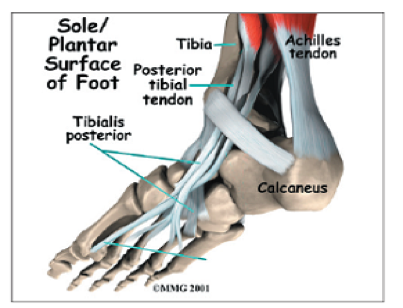
One of the most important tendons in the lower leg is the posterior tibial tendon. This tendon starts in the calf, stretches down behind the inside of the ankle and attaches to the bones in the middle of the foot. It helps hold up your arch and provides support as you step off on your toes when walking.
Causes
Progressive flatfoot often occurs in women over 50 years of age and may be due to an inherent abnormality of the tendon. There are several other risk factors, including:
- Obesity
- Diabetes
- Hypertension
- Previous surgery or trauma, such as an ankle fracture on the inner side of the foot
- Local steroid injections
- Inflammatory diseases such as Reiter's syndrome, rheumatoid arthritis, spondylosing arthropathy and psoriasis
- Athletes who are involved in sports such as basketball, tennis, soccer or hockey may tear the posterior tibial tendon. The tendon may also become inflamed if excessive force is placed on the foot, such as when running on a banked track or road.
Diagnosis
Diagnosis is based on a physical examination and history. Your physician may ask you to stand on your bare feet facing away, to view how your foot functions. You may also be asked to stand on your toes or to do a single heel rise. With your hands placed on a wall, you will lift the unaffected foot off the ground and rise up on the toes of the other foot. Normally, the heel will rotate inward; the absence of this sign indicates posterior tibial tendon dysfunction. Your doctor may request X-rays, an ultrasound or an MRI of the foot.