
What is Ankle Arthroscopy?
Ankle arthroscopy is a procedure where a telescope is placed inside your ankle joint. The origins of the word are arthro (meaning joint) and scope (meaning a telescope). Through a small incision, a mini telescope is introduced into the joint to visualise the joint line. Through a secondary incision, an instrument is introduced to remove inflammation, debris, and resect damage. One of the most common applications of ankle arthroscopy is in conjunction with ankle reconstruction surgery. Modern ankle reconstruction’s have a 91% success rate in the resolution of pain and the return to normal function.
What is Arthroscopy used for?
Arthroscopy can be used to diagnose and treat different disorders of the ankle joint. The goals of surgery are to reduce ankle pain and improve overall function. The list of problems that can be treated with this technology is constantly evolving and includes:
- Ankle arthritis: ankle fusion is a treatment option for many patients with end-stage ankle arthritis. Ankle arthroscopy offers a minimally invasive way to perform ankle fusion and the results can be equal to or better than open techniques.
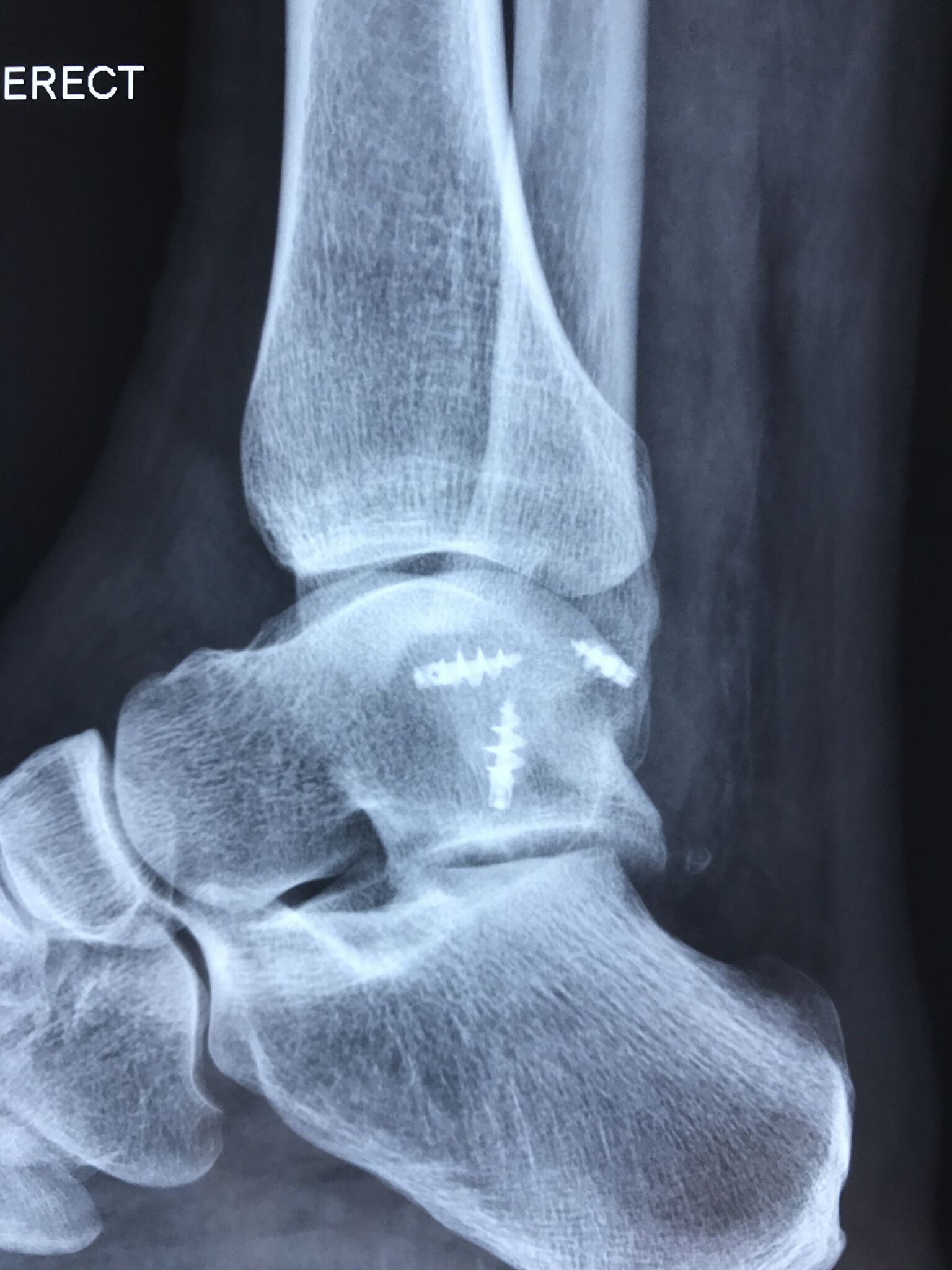
- Ankle fractures: arthroscopy may be used together with open techniques to repair fractures. It can assist with bone and cartilage alignment and can also be used to detect cartilage injuries within the ankle.
- Ankle instability: stretched ankle ligaments lead to a feeling that the ankle is giving way. The ligaments can be tightened and arthroscopic techniques may be an option.
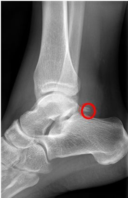
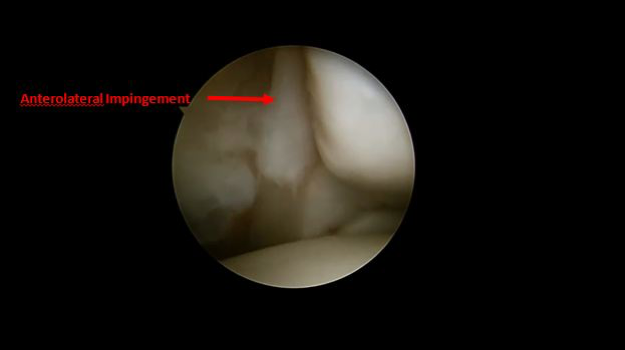
- Anterior ankle impingement (also referred to as athlete’s ankle or footballer’s ankle), this occurs when the bone or soft tissue at the front of the ankle joint becomes inflamed. Symptoms include ankle pain and swelling. Bending the ankle upward can become limited and uphill walking is often painful. Osteophytes (bone spurs) are revealed by X-ray. Arthroscopy is used to shave away inflamed tissues and bone spurs.
- Arthrofibrosis refers to the scar tissue that can form within the ankle which can lead to painful and stiff joints. Arthroscopy can be used to identify the scar tissue and remove it.
- Infection in the joint space cannot be treated with antibiotics alone. It often requires an urgent surgery to wash out the joint, which can be done with arthroscopy.
- Loose bodies: cartilage, bone and scar tissue can become free floating in the joint. Loose bodies can be painful and can cause problems such as clicking and catching and locking of the ankle joint. Ankle arthroscopy can be used to detect and remove loose bodies.
- Osteochondral defect (OCDs) are areas of damaged cartilage and bone in the ankle joint and are usually caused by fractures and sprains. Symptoms include ankle pain, swelling and catching or clicking in the ankle. Diagnosis is made with a combination of a physical exam and imaging studies including X-rays, MRI or CT scan. Treatment is based on the size, location and stability of the OCD and patient symptoms and activity demands. Surgery often consists of removal of the damaged cartilage and drilling small holes in the bone to promote healing. Bone grafting and cartilage transplant procedures may also be performed.
- Posterior ankle impingement: this overuse syndrome occurs when the soft tissue at the back of the ankle becomes inflamed. It's common amongst dancers can be associated with an extra bone called an os trigonum. The problem tissue can be removed with arthroscopy.
- Synovitis: the soft tissue lining of the ankle joint (synovial tissue) can become inflamed leading to pain and swelling. Causes include injury, overuse, rheumatoid arthritis and osteoarthritis. Ankle arthroscopy can be used to surgically remove inflamed tissue that does not respond to nonsurgical treatment.
- Unexplained ankle symptoms: occasionally, patients develop symptoms that cannot be explained by other diagnostic techniques. Arthroscopy provides the opportunity to look directly into the joint.